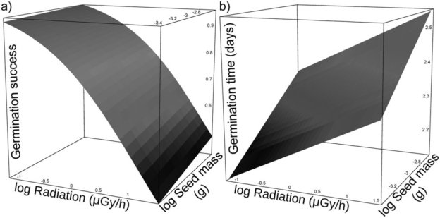
Ionizing radiation from Chernobyl affects development of wild carrot plants. Abstract
Latest Chernobyl paper shows radiation effects of wild carrots!
Abstract
“Radioactivity released from disasters like Chernobyl and Fukushima is a global hazard and a threat to exposed biota. To minimize the deleterious effects of stressors organisms adopt various strategies. Plants, for example, may delay germination or stay dormant during stressful periods. However, an intense stress may halt germination or heavily affect various developmental stages and select for life history changes. Here, we test for the consequence of exposure to ionizing radiation on plant development. We conducted a common garden experiment in an uncontaminated greenhouse using 660 seeds originating from 33 wild carrots (Daucus carota) collected near the Chernobyl nuclear power plant. These maternal plants had been exposed to radiation levels that varied by three orders of magnitude. We found strong negative effects of elevated radiation on the timing and rates of seed germination. In addition, later stages of development and the timing of emergence of consecutive leaves were delayed by exposure to radiation. We hypothesize that low quality of resources stored in seeds, damaged DNA, or both, delayed development and halted germination of seeds from plants exposed to elevated levels of ionizing radiation. We propose that high levels of spatial heterogeneity in background radiation may hamper adaptive life history responses.”
Zbyszek Boratyński, Javi Miranda Arias, Cristina Garcia, Tapio Mappes, Timothy A. Mousseau, Anders P. Møller, Antonio Jesús Muñoz Pajares, Marcin Piwczyński & Eugene Tukalenko
Ionizing Radiation from Chernobyl and the Fraction of Viable Pollen
Tim Mousseau – latest Chernobyl paper in International Journal of Plant Sciences:
Oct 05, 2016
Pollen viability is an important component of reproductive success, with inviable pollen causing failure of reproduction. Pollen grains have evolved mechanisms to avoid negative impacts of adverse environmental conditions on viability, including the ability to sustain ionizing radiation and repair DNA. We assessed the viability of 109,000 pollen grains representing 675 pollen samples from 111 species of plants in Chernobyl across radiation gradients that spanned three orders of magnitude. We found a statistically significant but small and negative main effect of radiation on pollen viability rates across species (Pearson’s r = 0.20). Ploidy level and the number of nucleate cells (two vs. three) were the only variables that influenced the strength of the effect of radiation on pollen viability, as reflected by significant interactions between these two variables and background radiation, while there were no significant effects of genome size, pollen aperture type, life cycle duration, or pollination agent on the strength of the effect of radiation on pollen viability.
Introduction
Most organisms are susceptible to environmental perturbations—such as climate change, extreme weather events, pollution, changes in nutrient availability, and changes in ionizing radiation levels—but the effects of such perturbations on individuals, populations, and ecosystems are variable (Candolin and Wong 2012; IPCC 2013; Møller and Mousseau 2013). In order to better understand these effects and to predict how a given species would respond to environmental disturbances, a study of the specific effects at different stages of organisms’ life cycles is required. Since reproduction is a key phase in the life cycle of any organism, reproductive effects are of particular interest. In the case of the effects of ionizing radiation, the negative consequences for reproduction in response to acute irradiation have been studied for decades and are well established (review in Møller and Mousseau 2013). However, the effects of long-term chronic exposure to low dose radiation are poorly understood.
Pollen grains are susceptible to the effects of environmental perturbations, which can have significant negative consequences for plant reproduction through pollen limitation (Delph et al. 1997; Ashman et al. 2004). Potential negative environmental effects include those resulting from elevated levels of ionizing radiation (Koller 1943). Therefore, plants have mechanisms to protect themselves from such effects, such as DNA repair, bi- or trinucleate cells, or redundancies in the genome resulting from duplications.
The area around Chernobyl in Ukraine has proven particularly useful for studying the effects of radioactive contamination on ecological and evolutionary processes at a large spatial scale. The Chernobyl nuclear accident in April 1986 led to the release of between 9.35 × 10³ and 1.25 × 10⁴ petabecquerel of radionuclides into the atmosphere (Møller and Mousseau 2006; Yablokov et al. 2009; Evangeliou et al. 2015). These radioactive contaminants were subsequently deposited in the surrounding areas of Belarus, Russia, and Ukraine but also elsewhere across Europe and even in Asia and North America. The pattern of contamination is highly heterogeneous, with some regions having received much higher levels of radionuclides than others, owing to atmospheric conditions at the time of the accident (fig. 1). To this day, the Chernobyl area provides a patchwork of sites that can differ in radioactive contamination level by up to five orders of magnitude across a comparatively small area. Even decades after the accident, the amount of radioactive material remaining around Chernobyl is enormous (Møller and Mousseau 2006; Yablokov et al. 2009).
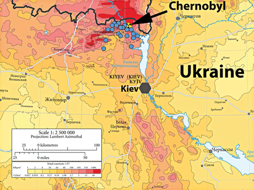
Fig. 1. Map of the distribution of radioactive contamination in the Chernobyl region, with pollen sampling locations marked. Adapted from DeCort et al. (1998).
Because of the unprecedented scale and global impact of the Chernobyl event, it is not surprising that it generated significant interest in both the scientific community and the general public. As a result, studies have been conducted to assess the consequences of Chernobyl for human health and agriculture as well as its biological effects, ranging from the level of DNA to entire ecosystems. Since ionizing radiation has long been well established as a mutagen (Nadson and Philippov 1925; Muller 1950), a large proportion of the research effort has focused on examining changes in mutation rates in areas that have been radioactively contaminated to different degrees as a result of the accident. Although there is considerable heterogeneity in the results of these studies, most have detected significant increases in mutation rates or genetic damage following the Chernobyl disaster, with the rates remaining elevated over the following 2 decades (reviewed in Møller and Mousseau 2006). For example, the mean frequency of mutations in Scots pine (Pinus sylvestris) is positively correlated with the level of background radiation, and it is 10 times higher in contaminated areas compared with control sites (Shevchenko et al. 1996). A study of Scots pine seeds detected elevated mutation rates within the exclusion zone over a period of 8 yr following the accident (Kal’chenko et al. 1995). In wheat (Triticum aestivum), the mutation rate was six times higher in radioactively contaminated areas compared with controls (Kovalchuk et al. 2000). Likewise, the frequency of chromosomal aberrations in two varieties of wheat grown within the Chernobyl exclusion zone 13 yr after the disaster was elevated compared with the spontaneous frequency of chromosomal aberrations in these cultivars (Yakimchuk et al. 2001). The levels of chromosome aberrations in onions (Allium cepa) were also positively correlated with the intensity of radioactive contamination in plants grown 20 yr after the accident (Grodzinsky 2006). Therefore, there is considerable evidence showing increased mutation rates in plants in the most contaminated sites (Møller and Mousseau 2015).
On the basis of the results of these studies, one might expect that a similar relationship between radiation level and the frequency of abnormalities would be seen in pollen. Indeed, Kordium and Sidorenko (1997) reported that the frequency of meiotic anomalies in microspore formation and the frequency of pollen grain viability was reduced in 8%–10% of the 94 plant species studied as a function of the intensity of gamma radiation 6–8 yr after the accident. In violets (Viola matutina), the proportion of viable pollen was negatively correlated with background radioactive contamination (Popova et al. 1991). While it is evident that plants differ in their susceptibility to ionizing radiation, the reasons for this variation are not entirely clear. It is likely that some species develop tolerance and/or resistance to mutagenic effects of radiation to a greater extent than others (Baer et al. 2007). For example, pollen of silver birch (Betula verrucosa), which grows in areas contaminated by the Chernobyl accident, showed elevated DNA repair ability compared with pollen from control areas, consistent with adaptation or epigenetic responses to increased radiation (Boubriak et al. 2008). There are also indications that genome size might affect the response of different species to radiation. Among the plants studied by Kordium and Sidorenko (1997), the rate of pollen viability decreased with increasing radiation to a higher degree in plants with smaller genomes (Barnier 2005), although the actual mechanism remains unknown. One potential explanation is that a larger genome might contain multiple copies of some genes as a result of duplication, rendering mutations in one of these copies less deleterious than if there were only a single copy present, although this explanation may not universally apply (Otto 2003).
In order to assess the effects of radioactive contamination on plant reproduction and to further assess species-specific differences in the effects of ionizing radiation on pollen viability, we analyzed pollen samples from plants growing in the Chernobyl region. We expected that the effects of radiation would differ among species, with some plants showing higher pollen inviability rates than others as a result of elevated radiation levels. A second objective was to test whether observed differences in pollen viability rates could be attributed to differences in phenotype among species, with possible explanatory factors including pollen size, the number of pollen apertures, ploidy, genome size, bi- or trinucleate cells, life span (annual vs. perennial), and pollination agent. We hypothesized that each of these factors could be related to the plants’ ability to resist or to tolerate radiation-induced mutations. Pollen size, genome size, and ploidy are all related to the amount of DNA and the number of copies of genes contained in the pollen grain. Because the pollen aperture—as the site of pollen germination—could be particularly susceptible to radiation-induced damage, we included the number of apertures as a potential explanatory variable. Furthermore, whether a plant is annual or perennial is related to individual longevity and, consequently, to the number of mutations that can accumulate over its lifetime as well as to the number of generations from the time of the Chernobyl accident until the time of sample collection. This may be particularly relevant for plants, given that germ tissue is derived from somatic tissues during each reproductive event as opposed to most animals, in which germ cells terminally differentiate very early during embryonic development (Buss 2006). Pollen viability depends on the ability of pollen to assess the integrity of its DNA and to repair the DNA of the generative nuclei before division (Jackson and Linskens 1980). This process is particularly important for binucleate pollen cells in which this happens during pollen germination, which is in contrast to trinucleate pollen cells, in which the need for DNA repair during pollen germination is less evident. DNA repair efficiency and adaptation of plants to chronic irradiation may also depend on the composition of radiation at the contaminated sites (Boubriak et al. 1992, 2008).
Across all plant species, we found a statistically significant relationship between radiation and the frequency of viable pollen of an intermediate magnitude (Cohen 1988). We also documented significant interactions between species and radiation, radiation and cell number, and radiation and ploidy. However, the significant effect of ploidy disappeared when both ploidy and whether cells were bi- or trinucleate were entered simultaneously in a single model. Most effects were small to intermediate in magnitude, as is commonly the case in studies of living organisms (Møller and Jennions 2002). We emphasize that our study included by far the largest sample size so far reported to detect effects of chronic radiation on pollen viability. However, we also emphasize the limits of our study. Many plant species could not be included simply because we could not locate multiple flowering specimens during our fieldwork. These and other sampling limitations reduced the number of pollen grains and the number of species that could be included.
Species differ in their susceptibility to radiation, as demonstrated for birds at both Chernobyl and Fukushima (Møller and Mousseau 2007; Møller et al. 2013; Galván et al. 2014), and in terms of adaptation to radiation (Galván et al. 2014; Møller and Mousseau 2016; Ruiz-González et al. 2016). The observed interspecific differences in radiation effects reported here for the proportion of viable pollen could be due to adaptation to radiation through tolerance of radiation-induced mutations or through induction of increased DNA repair in organisms living in contaminated areas. Another possibility is that some species are more resistant to radiation because of historical exposure in radiation hotspot areas with high natural levels of radiation (Møller and Mousseau 2013).
We observed a significant relationship between the proportion of viable pollen and the interaction between ploidy and radiation. Such a finding might suggest that resistance to deleterious effects of radiation is based on redundancy in the genome, where species with higher ploidy levels have an advantage if they have multiple copies of a given gene. We failed to detect an effect of selected physical attributes of pollen grains—such as genome size, pollen size, and aperture type—on the susceptibility of pollen to radiation. Furthermore, whether a plant was annual or perennial or whether it was insect or wind pollinated did not affect the proportion of viable pollen. Finally, whether plants produced bi- or trinucleate pollen had a significant effect on pollen viability, and the interaction between radiation and cell number was also significant.
While we confirmed the general finding of Kordium and Sidorenko (1997) that in approximately 10% of species the proportion of viable pollen is negatively correlated with radiation level, we were unable to reproduce their findings with respect to the overall magnitude of this effect. Our observed effect size was much smaller, and the slopes for individual species differed significantly from those reported by Kordium and Sidorenko (1997). Because more than 10 yr have passed between the two studies, we suggest that a change in radiation effects has taken place over time, for example, as a result of adaptation or accumulation of mutations. Another possible explanation for the discrepancy has to do with sample size, since our study included a much larger number of pollen samples and sampling locations than the study by Kordium and Sidorenko (1997). These explanations are not necessarily mutually exclusive.
Whereas other studies have demonstrated significant negative effects of radioactive contamination around Chernobyl on mutation rates and fitness in general, our study of pollen viability shows a very small effect, and some species even show positive relationships between pollen viability and radiation that is suggestive of adaptation to increased levels of radiation. However, on the basis of the current study, it is not possible to determine whether the observed heterogeneity reflects evolved adaptive responses or is the consequence of unmeasured selective effects on characters correlated with pollen viability, which could in part explain an overall positive effect of radiation (for a discussion of evolutionary responses in Chernobyl, see Møller and Mousseau 2016). Experimental approaches would be needed to decipher the mechanisms underlying the heterogeneity in plant responses observed here (Mousseau 2000).
The observed variability in susceptibility to radiation is a common finding in studies of the effects of radiation from Chernobyl (Møller and Mousseau 2007; Galván et al. 2011, 2014; Møller et al. 2013). While our results are consistent with earlier findings that DNA repair mechanisms may play an important role in adaptation to life in radioactively contaminated environments—especially for plants, which are sessile and hence cannot move to less contaminated areas—further research is required to test this explicitly. Finally, because of the observed differences in resistance to radiation among species, it is likely that even small overall effects of radiation—such as the one on the proportion of viable pollen described here—can have significant consequences for species composition and abundance at a given location and, therefore, for ecosystem characteristics and functioning.
In conclusion, we have found a statistically significant overall negative relationship between radiation intensity and the frequency of viable pollen in plants growing in contaminated areas around Chernobyl. The magnitude of this effect across species included in our study was intermediate. We only found a significant relationship between the proportion of viable pollen and ploidy × radiation interaction, bi- or trinucleate cells, and bi- or trinucleate cells × radiation interaction. This suggests that DNA repair mechanisms could play an important role for the ability of plants to resist increased radiation, at least when it comes to pollen formation.
Acknowledgments
We thank Puri López-García for use of a microscope for pollen counts. This work has benefited from the facilities and expertise of the cytometry platform of Imagif (Centre de Recherche de Gif; http://www.imagif.cnrs.fr). We thank Spencer Brown and Mickaël Bourge for their help with the flow cytometry measurements and Srdan Randić for help with pollen counts. Field collections for this study were supported in part by the Centre National de la Recherche Scientifique (France), the North Atlantic Treaty Organization Collaborative Linkage Grant program, the Fulbright program, the University of South Carolina College of Arts and Sciences, and the Samuel Freeman Charitable Trust. Two reviewers provided constructive criticism.
Read full paper at:
http://www.journals.uchicago.edu/doi/full/10.1086/688873
No need for a nuclear reactor to produce medical isotopes: Canada shows the way.
14 September 2016. A consortium of institutions led by TRIUMF, Canada’s national laboratory for particle and nuclear physics and accelerator-based science, is granting sole rights for its proprietary technetium-99m (Tc-99m) production technology to ARTMS™ Products, Inc (ARTMS). Technetium-99m is used in over 80% of all nuclear medicine imaging procedures and is vital to patient care in areas such as cardiology, oncology, and neurology. …

Typically sourced from an ageing global reactor fleet, Tc-99m has been subject to significant supply disruptions in recent years. ARTMS’ production technology promises to provide a reliable, cost effective, and safe supply of this critical medical isotope. The license includes all the required products and procedures for the production of Tc-99m using common hospital-based and commercial cyclotrons, through the bombardment of a high-energy proton beam against specific chemical ‘targets’. ….
“The ARTMS production technology offers many advantages, and that is why we believe our technology is truly disruptive and that it will gain widespread adoption,” Dr. Schaffer added. “Not only does the ARTMS production technology provide regional supply security of Tc-99m, it also offers favourable economics, and aids to eliminate the need for highly-enriched uranium, which is currently used by nuclear reactors to produce this isotope.”
“This agreement represents the culmination of six years of hard work by a dedicated team from across Canada, including TRIUMF, the BC Cancer Agency, Lawson Health Research Institute, and the Centre for Probe Development and Commercialization,” said Dr. Jonathan Bagger, Director of TRIUMF. “Today marks the completion of a major milestone as we move to commercialize a decentralized, green, and Canadian-made, technology that can produce Tc-99m daily at hundreds of hospital-based cyclotrons around the world. This licensing agreement marks the beginning of a new era in Tc-99m production and supply security.”
More information on the recent global isotope shortages, Tc-99m, and the story of ARTMS can be found in this media backgrounder and more information on medical isotopes and cyclotrons can be found in this FAQ. http://www.triumf.ca/current-events/artms%E2%84%A2-products-inc-licenses-canadian-technology-address-global-medical-isotope
Study pinpoints protein that detects damage from radiation
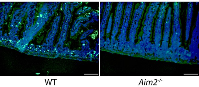
Small intestine tissue from mouse after high-dose X-ray radiation. Green fluorescence shows dying epithelial cells.
High doses of radiation from cancer treatment can cause severe damage to cells and tissues, resulting in injury to bone marrow and the gastrointestinal tract. The consequences can be fatal. Yet researchers do not fully understand how exposure to radiation triggers this damage at the molecular level.
Led by Yale professor of immunobiology Richard Flavell, an international team of researchers studied the radiation response using animal models. They identified a novel mechanism of radiation-induced tissue injury involving a protein called AIM2, which can sense double-strand DNA damage and mediate a special form of cell death known as pyroptosis.
They observed that in animals lacking AIM2, both the gastrointestinal tract and bone marrow were protected from radiation. While the role of AIM2 as a sensor that detects infectious threats to the body was known, this study is the first to describe the protein’s function in the detection of radiation damage to the chromosomes in the nucleus, said the researchers.
When a cell receives a high dose of radiation, the DNA is broken into pieces, which can be joined together again. However this aberrant rejoining of chromosomal fragments can lead to chromosomal abnormalities and cancer. Flavell and his team believe that when this chromosomal damage is inflicted, the AIM2 pathway is activated in order to kill the cell to avoid the deleterious consequences of these chromosomal translocations, such as those commonly seen in cancer cells.
For this reason, the cells that accumulate this chromosomal damage are dangerous to the person or animal and are therefore killed by this AIM2 pathway. This pathway is beneficial to the person or animal under normal circumstances because it eliminates dangerous cells, but when a high dose of radiation is given the pathway is detrimental because it leads to bone marrow and digestive tract injury.
These findings suggest that a drug that blocks or inhibits the AIM2 pathway could potentially limit the deleterious side effect of chemotherapy or radiotherapy on cancer patients, said the researchers.
Read the full paper in Science.
http://news.yale.edu/2016/11/10/study-pinpoints-protein-detects-damage-radiation
Russia, Japan Team Up to Study How Radiation Affects the Next Generation’s DNA
Russia and Japan are set to team up to become leaders in transgenerational healthcare research, to help prevent the effects of nuclear catastrophes being passed genetically from one generation to the next indefinitely.
Both Russia and Japan have a stake in this research, given that both countries are still dealing with radiation exposure via the events in Nagasaki, Hiroshima, Fukushima and Chernobyl. “This research is extremely important in relation to future generations we are responsible for,” said Nomura Taisei, Radiation Biology and Medical Genetics Department Head at National Institute for Biomedical Research at Osaka University.
The professor was at the 15th Congress on Innovation Technologies in Pediatrics and Pediatric Surgery which was held in Moscow from October 25-27, making a report on trasngenerational healthcare. His report shines a light on how exposure to radiation is passed down through generations via DNA mutation.
When DNA is damaged, the consequences for future generations are serious.Birth abnormalities, developmental disorders, a weakened immune system, higher cancer risks, and numerous physical and mental disorders are all the result of these gene mutations passed down to future generations. While the effects of radiation exposure passing between generations has so far not been widely studied in humans, the effects on experimental animal subjects is more widely understood.
Professor Nomura’s experiments on mice proved that genetic effects of radiation exposure can cause genetic defects into the 58th generation. The problem is that Japan has very little data on radiation exposure on humans.
This is where Russia can help, through opening up their database on three tree generations of people: those who were exposed after the Chernobyl disaster, those who were exposed prenatally, and those whose parents were exposed before impregnation. Thus Russia and Japan can now conduct joint comparative research of the effects of radiation on animals and on humans applying the latest technologies.
The Head of Children’s Scientific and Practical Center of Radiation Protection, Larisa Naleva told Sputnik Japan about the importance of this Russian-Japanese research project.
“We assume that the phenomenon of radiation-induced genetic instability has significant effects not only on the health of exposed people but also on the health of their children, first of all, resulting in an increased cancer risk. We have already detected an increase of morbidity in the second generation of exposed people’s descendants and now we are studying the third generation. Today in Russia there are about 135 thousand children who have been exposed or are exposed to radiation to some extent,” said Naleva. By using Japan’s expertise, Naleva hopes that the health risk for subsequent generations of those who were exposed to radiation can be reduced. “And that is the goal of our collaboration with our Japanese colleagues,” she said.
https://sputniknews.com/society/201611021046998030-russia-japan-radiation-dna/
Mother’s Radiation Lab and Clinic
The relatively short video shows a female perspective of how women are dealing with the risk despite the Japanese governments, lack of radiation testing, children’s health checks, financial and social support – the social responsibility to their community
Women suffer the most from this stoic denial that radiation effects the community, causing unnecessary stress from risk of radionuclide ingestion on a child’s growing body, well established to be many times more sensitive to radiation due to rapidly dividing cells programmed by DNA at risk during early development
It is sad a mother’s worldview has been largely left out of the South Australian debate around the whole nuclear cycle dominated by senior male nuclear sales executives and academics
However, that isn’t any surprise, as that is how the world embraced the whole nuclear industry in the first place, that is from a purely patriarchal worldview and that is a matter of our species shameful human history https://www.facebook.com/groups/1314655315214929/
Ionizing radiation May Contribute to Development of Alzheimer’s

University of Southern Denmark
More humans than ever are exposed to higher levels of ionizing radiation from medical equipment, airplanes, etc. A new study suggests that this kind of radiation may be a confounding factor in the neurodegenerative disease Alzheimer´s.
Alzheimer’s disease is the leading cause for dementia in the elderly, and its global prevalence is supposed to increase dramatically in the following decade – up to 80 million patients by 2040.
– It is crucial that we investigate the potential factors behind this disease, says postdoc Stefan J. Kempf, University of Southern Denmark. His research focuses on possible connections between radiation and cognitive impairments.
In a new study, he and an international consortia involving colleagues from Italy, Japan, Germany and Denmark show that low doses of ionising radiation induce molecular changes in the brain that resemble the pathologies of Alzheimer’s.
The study has been published in Oncotarget. Co-authors are from Institute of Radiation Biology/Institute of Pathology, Helmholtz Zentrum München, German Research Center for Environmental Health and Institute for Environmental Sciences in Japan.
Large numbers of people of all age groups are increasingly exposed to ionizing radiation from various sources. Many receive chronic occupational exposure from nuclear technologies or airline travel. The use of medical diagnostics and therapeutic radiology has increased rapidly – for example more than 62 million CT scans per year are currently carried out in USA.
Approximately one third of all diagnostic CT examinations are scans of the head region.
– All these kinds of exposures are low dose and as long as we talk about one or a few exposures in a lifetime I do not see cause for concern. What concerns me is that modern people may be exposed several times in their lifetime and that we don’t know enough about the consequences of accumulated doses, says Stefan J. Kempf.
Recent data suggest that even relatively low radiation doses, similar to those received from a few CT scans, could trigger molecular changes associated with cognitive dysfunction.
In their new study, the researchers have elucidated molecular alterations in the hippocampus of mice. The hippocampus is an important brain region responsible for learning and memory formation and it is known to be negatively affected in Alzheimer´s.
The authors induced changes in the hippocampus by two kinds of chronic low-dose-rate ionizing radiation treatments. The mice were exposed to cumulative doses of 0.3 Gy or 6.0 Gy given at low dose rates of 1 mGy over 24 hours or 20 mGy over 24 hours for 300 days.
– Both dose rates are capable of inducing molecular features that are reminiscent of those found in the Alzheimer’s disease neuropathology, says Stefan J. Kempf.
When a patient gets a head scan, the doses varies between 20 and 100 mGy and lasts for around one minute. When a person flies, he or she gets exposure to ionising radiation coming from space but the rates are by far smaller than a CT scan.
– When you compare these figures you will find that we exposed the mice to a more than 1000 times smaller cumulative dose than what a patient gets from a single CT scan in the same time interval. And even then we could see changes in the synapses within the hippocampus that resemble Alzheimer´s pathology.
According to Stefan J. Kempf, the data indicate that chronic low-dose-rate radiation targets the integration of newborn neurons in existing synaptic wires.
###
Paper: Chronic low-dose-rate ionising radiation affects the hippocampal phosphoproteome in the ApoE?/? Alzheimer mouse model. Forfattere: Stefan Kempf, Dirk Janik, Zarko Barjaktarovic, Ignacia Braga-Tanaka III, Satoshi Tanaka, Frauke Neff, Anna Saran, Martin Røssel Larsen, Soile Tapio. OncoTarget, 20. september 2016.
https://www.eurekalert.org/pub_releases/2016-10/uosd-ctr102716.php
Causal connection between nuclear radiation and Alzheimers’ Disease – European research
Concern that radiation may contribute to development of Alzheimer’s https://www.eurekalert.org/pub_releases/2016-10/uosd-ctr102716.php UNIVERSITY OF SOUTHERN DENMARK MORE HUMANS THAN EVER ARE EXPOSED TO HIGHER LEVELS OF IONIZING RADIATION FROM MEDICAL EQUIPMENT, AIRPLANES, ETC. A NEW STUDY SUGGESTS THAT THIS KIND OF RADIATION MAY BE A CONFOUNDING FACTOR IN THE NEURODEGENERATIVE DISEASE ALZHEIMER´S.
Alzheimer’s disease is the leading cause for dementia in the elderly, and its global prevalence is supposed to increase dramatically in the following decade – up to 80 million patients by 2040.
– It is crucial that we investigate the potential factors behind this disease, says postdoc Stefan J. Kempf, University of Southern Denmark. His research focuses on possible connections between radiation and cognitive impairments.
In a new study, he and an international consortia involving colleagues from Italy, Japan, Germany and Denmark show that low doses of ionising radiation induce molecular changes in the brain that resemble the pathologies of Alzheimer’s. Continue reading
Radiation-absorption tests under development could save lives in nuclear explosion
Direct measurement (like Becquerels) via blood samples described in the article sounds like the way to go.
“The key to understand is that this is something that has never existed and we hope it never gets used,” Josh LaBaer, principal investigator and director of the Biodesign Institute at Arizona State University, told Homeland Preparedness News.
The tests could also have civilian applications as well, LaBaer said, such as in the event of industrial accidents at a nuclear power plant or in medical situations when people are exposed to excessive radiation.

The U.S. government is funding the late-stage development of tests that would quickly determine how much radiation a person has absorbed in the event of a catastrophic nuclear explosion.
The U.S. Department of Health and Human Services’ Office of the Assistant Secretary for Preparedness and Response (ASPR) is sponsoring the development of tests that go beyond detecting whether radiation is on a person’s skin to determining the amount of radiation that has been absorbed into a person’s body.
“The key to understand is that this is something that has never existed and we hope it never gets used,” Josh LaBaer, principal investigator and director of the Biodesign Institute at Arizona State University, told Homeland Preparedness News.
ASPR’s Biomedical Advanced Research and Development Authority (BARDA) will provide more than $21.3 million over four years to develop the tests. Kansas City, Missouri-based MRIGlobal said in a written statement the contract could be extended for up to $100 million over 10 years.
MRIGlobal is partnering with Thermo Fisher Scientific and Arizona State University to lead the development of the program for BARDA. The agency also will provide more than $22.4 million in funding over two years to DxTerity Diagnostics based near Los Angeles.
“The challenge was that in the event of a nuclear bomb in a major American city, there is an instantaneous release of high doses of gamma radiation, which is the type of radiation that travels through the air over large distances,” LaBaer said. “In that type of mass casualty event there would be lots of people who would need to be evaluated.”
The task for researchers was to develop a device that could quickly measure how much radiation large numbers of people had potentially absorbed into their organs and blood cells during a nuclear emergency. Devices currently available today can only detect radiation on the skin.
“The amount of radiation that gets absorbed into the body has a direct implication on how that person gets triaged and managed,” LaBaer said. Absorption of a small or moderate dose of radiation could require medication, while a larger dose could require hospitalization and a potential bone marrow transplant.
BARDA is supporting development of the tests with the goal of potentially purchasing them from one or more of the companies for the Strategic National Stockpile.
After a six-year effort, the university has developed the ASU radiation (ARad) biodosimetry test, which would generate results in about eight hours and could be used on people who were exposed to radiation up to seven days after the event. HHS said the potential exists where 400,000 or more tests could be processed a week.
In the test, a blood sample is taken to isolate the white blood cells in order to collect the genes that have been exposed to radiation. Certain genes are more predictive when it comes to determining the amount of radiation the body was exposed to.
“We were looking for the smallest number of genes we could use but that still were accurate in predicting dose depending on the time after the event,” LaBaer said.
Work to date has been based on animal studies and developing conversion factors to transfer to humans.
The tests could also have civilian applications as well, LaBaer said, such as in the event of industrial accidents at a nuclear power plant or in medical situations when people are exposed to excessive radiation.
Admiral Rickover “father” of the U.S. nuclear navy called for outlawing nuclear reactors
 Bailing out aging nuclear power plants can impact development of renewable energy technologies, Enformable, 17 Oct 2016 “………Rickover: “Outlaw Nuclear Reactors”
The bottom line when it comes to nuclear power comes from Admiral Rickover, considered the “father” of the U.S. nuclear navy as well as being in charge of building Shippingport. When he retired from the Navy in 1982 he addressed a Congressional committee and said—his remarks are included in Cover Up—that until several billion years ago “it was impossible to have any life on Earth; that is, there was so much radiation on Earth you couldn’t have any life—fish or anything. “ Then, “gradually, “the amount of radiation on this planet and probably in the entire system reduced and made it possible for some form of life to begin.”
“Now,” he went on, by utilizing nuclear power “we are creating something which nature tried to destroy to make life possible…every time you produce radiation,” a “horrible force” is unleashed, “in some cases for billions of years.” In other words, nuclear power plants recreate the very radioactive poisons that precluded life from existing. “And,” said Rickover, “I think there the human race is going to wreck itself.”
We must, for the sake of life, Rickover told the Congressional committee, “outlaw nuclear reactors.”
Rickover, deeply involved in nuclear technology, finally saw—as we all must—the light.http://enformable.com/
If they didn’t have mental problems before, Mars travellers sure will afterwards!
 Mars-goers may face permanent brain damage from cosmic radiation Oct. 12, 2016 Deep space travel could cause serious, irreversible brain damage, NBC News reports. Scientists have long known that leaving Earth’s magnetosphere—the magnetic bubble of plasma surrounding our planet—strips astronauts of their protection from radioactive particles, putting them at higher risk for health issues, including heart disease. Now, a new study out this week in Scientific Reports suggests that changes at the cellular level could also lead to worsened anxiety and even brain cancer. That could be bad news for NASA and other commercial space companies that want to send humans to the Red Planet by 2030. But NASA is working on it: The agency is researching methods to prevent exposure to radiation, which could find their way into new, improved space suits. http://www.sciencemag.org/news/sifter/mars-goers-may-face-permanent-brain-damage-cosmic-radiation
France: Public consultation on the draft decree on protection against the dangers of ionizing radiation
Sean Arclight Hervé Courtois CRIIRAD calls to mobilize against the adoption of very high reference levels
to manage nuclear accidents and their consequences.
The French authorities are preparing to establish the zones management criteria contaminated following a nuclear accident (or after an attack affecting a nuclear installation). What level of radiation exposure, and thus risk, will be taken in reference to decide whether or not to hire a particular action to protect the population? Very concretely: to what level of risk you will be condemned to live in contaminated areas? At what level of risk can you expect to be compensated and rehoused in a healthy environment?
The French authorities have retained the levels of effective dose as high as possible: 100 mSv for the accident phase and 20 mSv / year for accidental post phase (while for the public, the maximum dose limit is typically 1 mSv / year and that this value is already at a high level of risk). More limitations are high, lower are the expenses related to the protection and compensation for damage. This choice is unfortunately consistent with the capping of compensation for victims of a major nuclear accident. Nuclear power is exempted from the application of the polluter pays principle: they are the victims who bear the health and economic consequences of the disaster.
This decision does not just happen. It is the fruit of 20 years of efforts of the nuclear lobby, and specifically the French nuclear lobby via the Trojan horse, the FNEC (1). The key idea is to convince people that can be done entirely live in contaminated areas. Just a bit of training and equipment to control their environment, food. These experts have just “forgot” the central problem of the deteriorating health status of people, especially children.
If you are shocked by the image of the Japanese children wear around their necks a dosimeter as a pendant, if it is not the future you want for your children, act!
1. Study Centre on the Protection of the evaluation in the field Nuclear: an association with 4 members (EDF, AREVA, CEA and IRSN) and has widely infiltrated the national and international decision-making and including the ICRP (Jacques Lochard, Director of the FNEC, is now vice chairman of the main committee)
The Ministry of Environment, Energy and Marine has launched a consultation on the draft decree on the Directive 2013/59. Remember that France must transpose the Directive into French law by January 2018. This consultation is an opportunity to denounce the proposals which we find unacceptable and show already our requirements. We later learned of this consultation will end on 30 September.
Take part in the public consultation
and say NO to the obligation to live in contaminated areas!
> Learn more
> How to participate in the public consultation?
The Directive covers many topics which will be discussed further. Other actions will be implemented in the coming weeks. We already rely on your help to relay! https://www.facebook.com/groups/1021186047913052/
Mutational signatures of ionizing radiation in second malignancies
This article is important, and should be seen by as many people as possible, as this scientific study will impact greatly the future of our anti-nuclear cause.
By establishing the genetic signatures of any cancer caused by ionizing radiation, any future denial from the nuclear lobby is now impossible. Those scientifically established signatures will also be extremely helpful in court for any future suit from radiation victims.
Abstract
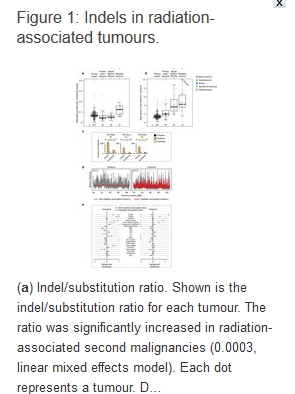
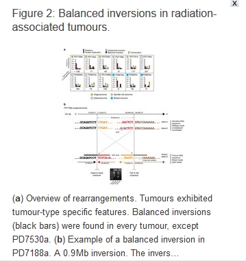
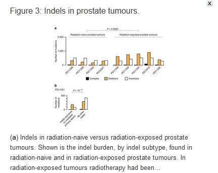
Ionizing radiation is a potent carcinogen, inducing cancer through DNA damage. The signatures of mutations arising in human tissues following in vivo exposure to ionizing radiation have not been documented. Here, we searched for signatures of ionizing radiation in 12 radiation-associated second malignancies of different tumour types. Two signatures of somatic mutation characterize ionizing radiation exposure irrespective of tumour type. Compared with 319 radiation-naive tumours, radiation-associated tumours carry a median extra 201 deletions genome-wide, sized 1–100 base pairs often with microhomology at the junction. Unlike deletions of radiation-naive tumours, these show no variation in density across the genome or correlation with sequence context, replication timing or chromatin structure. Furthermore, we observe a significant increase in balanced inversions in radiation-associated tumours. Both small deletions and inversions generate driver mutations. Thus, ionizing radiation generates distinctive mutational signatures that explain its carcinogenic potential.
Introduction
Exposure to ionizing radiation increases the risk of subsequent cancer. This risk exhibits a strong dose–response relationship, and there appear to be no safe limits for radiation exposure1. This association was first noted by March who observed an increased incidence of leukaemia amongst radiologists2. A leading cause of radiation-induced cancers appears to be exposure to medical radiation, either in the form of radiotherapy for an unrelated malignancy3 or diagnostic radiography4, 5. These iatrogenic tumours arise as de novo neoplasms in a field of therapeutic radiation after a latency period that can span decades6, and are not recurrences of the original cancer7.
Many, but not all, environmental carcinogens induce cancer by increasing the rate of mutation in somatic cells. The physicochemical properties of a given carcinogen govern its interaction with DNA, leading to recurrent ‘signatures’ or patterns of mutations in the genome. These can be reconstructed either from experimental model systems8, 9 or from statistical analyses of cancer genomes in exposed patients10, 11, 12. Ionizing radiation directly damages DNA, and can generate lesions on single bases, single-stranded nicks in the DNA backbone, clustered lesions at several nearby sites and double-stranded DNA breaks13. In experimental systems exposed to radiation, including the murine germline and Arabidopsis thaliana cells, ionizing radiation can cause all classes of mutations, with possible enrichment of indels14, 15, 16, 17, 18, 19, 20, 21, 22. Targeted gene screens in radiation-induced sarcoma have indicated an increased burden of deletions and substitutions with frequent inactivation of TP53 and RB1 (refs 23, 24, 25). In addition, a transcriptome profile that represents a state of chronic oxidative stress has been proposed to be specific to radiation-associated sarcoma26.
We studied the genomes of 12 radiation-associated second malignancies of four different tumour types: osteosarcoma; spindle cell sarcoma; angiosarcoma; breast cancer. These were secondary tumours that arose within a field of therapeutic ionizing radiation and were not thought to be recurrences of the original malignancy treated with radiation. We chose this experimental design for several reasons: the tumours are classic radiotherapy-induced cancers with high attributable risks for the radiation exposure; the radiation exposure occurs over a short time period relative to the evolution of the cancer; and the mutational signatures of sporadic breast cancers and sarcomas have been well documented10, 27, 28, 29. It should be noted that in the absence of biomarkers, a diagnosis of a tumour being radiation-induced cannot be definitively made (see Supplementary Note 1 for clinical details and further discussion).
We subjected these 12 tumours, along with normal tissues from the same patients, to whole-genome sequencing and obtained catalogues of somatic mutations. We compared our findings to 319 radiation-naive breast cancers and sarcomas processed by the same sequencing and bioinformatics pipeline: 251 breast tumours; 33 breast tumours with pathogenic BRCA1 or BRCA2 germline mutations; 35 osteosarcomas (see Methods for cohort details). In addition, we validated our findings in a published series of radiation-naïve and radiation-exposed prostate tumours from ten patients30.
The main aim of our analyses was to search for tumour-type independent, overarching signatures of ionizing radiation. Overall we identified two such signatures in radiation-associative second malignancies, an excess of balanced inversions and of small deletions.
To read more :
http://www.nature.com/ncomms/2016/160907/ncomms12605/full/ncomms12605.html
DNA damage, cancer caused by ionizing radiation identified
This UPI article was published on Sept. 13, 2016.
I added below the source of that UPI article, the study published on the sciences website “Nature” on Sept. 12, 2016.
This article is important, and should be seen by as many people as possible, as this scientific study will impact greatly the future of our anti-nuclear cause.
By establishing the genetic signatures of any cancer caused by ionizing radiation, any future denial from the nuclear lobby is now impossible. Those scientifically established signatures will also be extremely helpful in court for any future suit from radiation victims.
Researchers found mutational signatures left by radiation-caused changes to DNA, which may lead to better treatment of cancers.
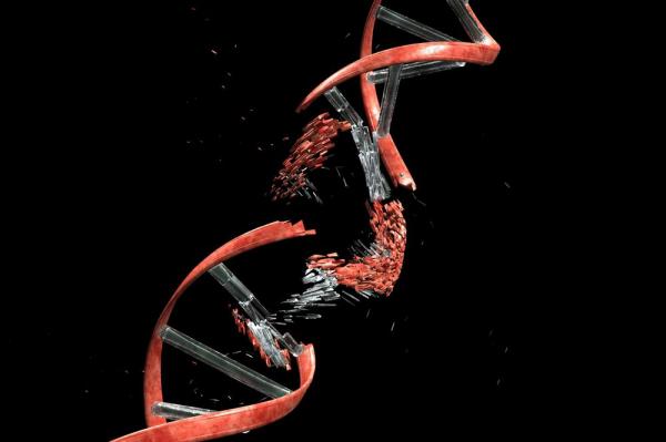
Researchers found mutational signatures which appear to indicate changes to DNA caused by exposure to ionizing radiation, which may allow doctors to better treat cancer caused by non-spontaneous mutations.
LONDON, Sept. 13 (UPI) — Though scientists have suspected ionizing radiation can cause cancer, experiments conducted in England are the first to show the damage it inflicts on DNA and may allow doctors to identify tumors caused by radiation.
In a study published in the journal Nature Communications, scientists showed the effects of gamma rays, X-rays and radioactive particles on DNA, deciphering patterns they think will help differentiate between spontaneous and radiation-caused tumors, allowing for better cancer treatment.
“To find out how radiation could cause cancer, we studied the genomes of cancers caused by radiation in comparison to tumors that arose spontaneously,” Dr. Peter Campbell, a researchers at the Wellcome Trust Sanger Institute, said in a press release. “By comparing the DNA sequences we found two mutational signatures for radiation damage that were independent of cancer type. We then checked the findings with prostate cancers that had or had not been exposed to radiation, and found the same two signatures again. These mutational signatures help us explain how high-energy radiation damages DNA.”
For the study, the researchers looked for mutational signatures in 12 cancer patients with radiation-associated second malignancies, and compared their tumors to 319 from patients not exposed to radiation.
The researchers found two mutational signatures they link to radiation. While one causes small deletions of DNA bases, the other — called a balanced inversion — includes two cuts to DNA, with the middle piece spinning around and rejoining in the opposite direction.
These mutations, especially balanced inversions, which do not happen naturally in the body, increase the potential for cancer to develop, the researchers say.
“This is the first time that scientists have been able to define the damage caused to DNA by ionising radiation,” said Adrienne Flanagan, a professor at the University College London Cancer Institute. “These mutational signatures could be a diagnosis tool for both individual cases, and for groups of cancers, and could help us find out which cancers are caused by radiation. Once we have better understanding of this, we can study whether they should be treated the same or differently to other cancers.”
Mutational signatures of ionizing radiation in second malignancies
« Ionizing radiation is a potent carcinogen, inducing cancer through DNA damage. The signatures of mutations arising in human tissues following in vivo exposure to ionizing radiation have not been documented. Here, we searched for signatures of ionizing radiation in 12 radiation-associated second malignancies of different tumour types. Two signatures of somatic mutation characterize ionizing radiation exposure irrespective of tumour type. Compared with 319 radiation-naive tumours, radiation-associated tumours carry a median extra 201 deletions genome-wide, sized 1–100 base pairs often with microhomology at the junction. Unlike deletions of radiation-naive tumours, these show no variation in density across the genome or correlation with sequence context, replication timing or chromatin structure. Furthermore, we observe a significant increase in balanced inversions in radiation-associated tumours. Both small deletions and inversions generate driver mutations. Thus, ionizing radiation generates distinctive mutational signatures that explain its carcinogenic potential. »
http://www.nature.com/ncomms/2016/160907/ncomms12605/full/ncomms12605.html
Linear No Threshold Theory (LNT) of ionising radiation is backed by new research
Gamma radiation at a human relevant low dose rate is genotoxic in mice, Anne Graupner, Dag M. Eide, Christine Instanes, Jill M. Andersen, Dag A. Brede, Stephen D. Dertinger, Ole C. Lind, Anicke Brandt-Kjelsen, Hans Bjerke, Brit Salbu, Deborah Oughton, Gunnar Brunborg & Ann K. Olsen Scientific Reports 6, Article number: 32977 September 21016
Abstract
Even today, 70 years after Hiroshima and accidents like in Chernobyl and Fukushima, we still have limited knowledge about the health effects of low dose rate (LDR) radiation. Despite their human relevance after occupational and accidental exposure, only few animal studies on the genotoxic effects of chronic LDR radiation have been performed. Selenium (Se) is involved in oxidative stress defence, protecting DNA and other biomolecules from reactive oxygen species (ROS). It is hypothesised that Se deficiency, as it occurs in several parts of the world, may aggravate harmful effects of ROS-inducing stressors such as ionising radiation.
We performed a study in the newly established LDR-facility Figaro on the combined effects of Se deprivation and LDR γ exposure in DNA repair knockout mice (Ogg1−/−) and control animals (Ogg1+/−). Genotoxic effects were seen after continuous radiation (1.4 mGy/h) for 45 days. Chromosomal damage (micronucleus), phenotypic mutations (Pig-a gene mutation of RBCCD24−) and DNA lesions (single strand breaks/alkali labile sites) were significantly increased in blood cells of irradiated animals, covering three types of genotoxic activity.
This study demonstrates that chronic LDR γ radiation is genotoxic in an exposure scenario realistic for humans, supporting the hypothesis that even LDR γ radiation may induce cancer……..

In the present study we demonstrate that exposure to a human relevant LDR γ radiation induces genotoxic effects in mouse blood cells assessed with three separate but complementary assays. These effects were expressed as increased levels of chromosomal damage (micronuclei), phenotypic mutations (RBCCD24−) and DNA lesions (ssb/als). The absolute measured changes were small, but significant. The formation of MN was observed in all irradiated groups independent of genotype or diet, and significant changes were seen in both immature and mature erythrocytes. This is an expected result given the chronic exposure and lack of splenic filtration of circulating MN-containing erythrocytes18……..In summary, exposure to chronic LDR of ionising radiation is indeed genotoxic with potential implications for cancer development, and the response is modified by the availability of Se, an element involved in the antioxidative defence report http://www.nature.com/articles/srep32977
1 This Month


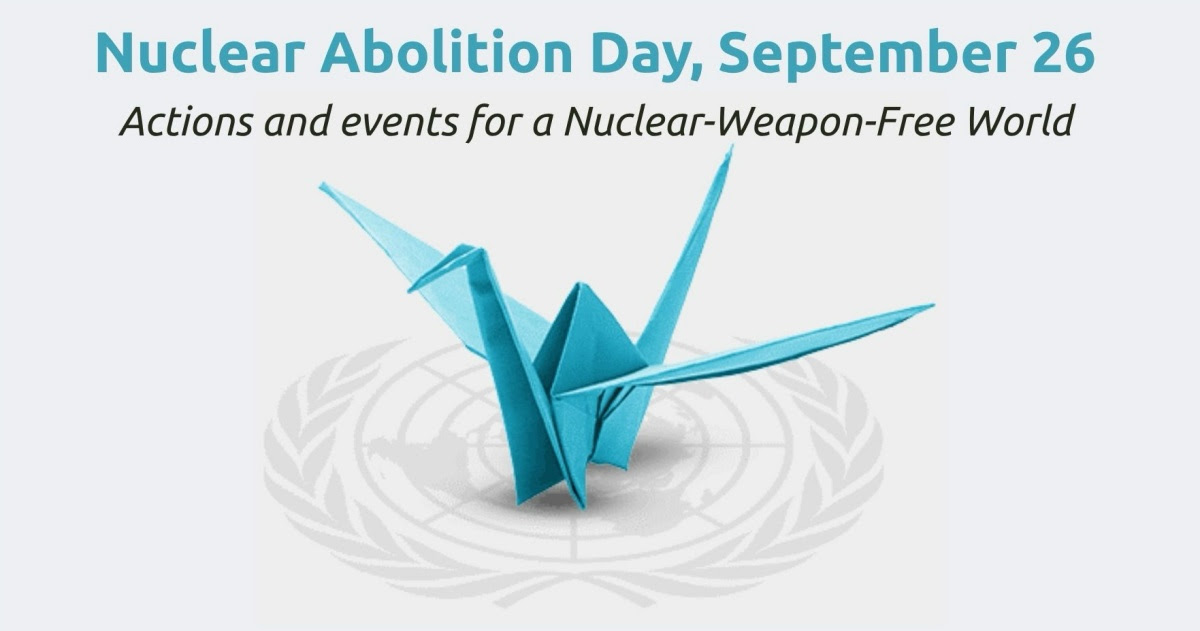
26 September – International Day for the Total Elimination of Nuclear Weapons

PLEASE OPPOSE HR 8255 AND S.3639 – bills would accelerate satellite deployment, with no environmental review or safeguards – https://ncstd5li.quorum.us/campaign/169331/
To see nuclear-related stories in greater depth and intensity – go to https://nuclearinformation.wordpress.com
-
Archives
- September 2026 (219)
- August 2026 (330)
- July 2026 (355)
- June 2026 (287)
- May 2026 (306)
- April 2026 (356)
- March 2026 (251)
- February 2026 (267)
- January 2026 (308)
- December 2025 (358)
- November 2025 (359)
- October 2025 (375)
-
Categories
- 1
- 1 NUCLEAR ISSUES
- business and costs
- climate change
- culture and arts
- ENERGY
- environment
- health
- history
- indigenous issues
- Legal
- marketing of nuclear
- media
- opposition to nuclear
- PERSONAL STORIES
- politics
- politics international
- Religion and ethics
- safety
- secrets,lies and civil liberties
- spinbuster
- technology
- Uranium
- wastes
- weapons and war
- Women
- 2 WORLD
- ACTION
- AFRICA
- Atrocities
- AUSTRALIA
- Christina's notes
- Christina's themes
- culture and arts
- Events
- Fuk 2022
- Fuk 2023
- Fukushima 2017
- Fukushima 2018
- fukushima 2019
- Fukushima 2020
- Fukushima 2021
- general
- global warming
- Humour (God we need it)
- Nuclear
- RARE EARTHS
- Reference
- resources – print
- Resources -audiovicual
- Weekly Newsletter
- World
- World Nuclear
- YouTube
-
RSS
Entries RSS
Comments RSS
 nuclear-news