Radiation damage to any cells but the reproductive organs. Genetic damage. Damage to the reproductive cells. Birth defects may result.
Genetic damage from radiation highlights need to protect physicians in cath lab, Cardiovascular Business, Jan 12, 2018 | Daniel AllarA pair of studies published in October added to the growing literature on the harmful effects of radiation exposure to interventional cardiologists in the cath lab.
One study showed brain-specific microRNA (miRNAs) was significantly down regulated in operators exposed to radiation when compared to age- and sex-matched controls who had no occupational exposure. Another found genetic biomarkers for DNA damage/repair rose significantly after procedures that required radiation but then returned to baseline levels after 24 hours. Notably, the use of protective leg shielding mitigated the damage.
Charles E. Chambers, MD, who authored an editorial accompanying the studies in Circulation, spoke to Cardiovascular Business about this research and recent developments that could improve operator safety. Here are three key takeaways from that conversation:
1. These studies were novel because they showed genetic damage related to radiation exposure.
This differs from previous research, Chambers said, which centered on hard clinical outcomes or the development of orthopedic injuries from years spent wearing heavy lead clothing to shield against radiation.
“But now these two studies suggest that radiation is what we were worried about all along. It really does increase genetic alterations,” he said.
Because dysregulated miRNA has been linked to certain forms of epilepsy, Alzheimer’s disease and brain cancers, the study authored by Andrea Borghini and colleagues raises the concern that radiation may also tie into cognitive impairment.
“We’re fortunate, I think, that there aren’t more tumors, there aren’t more long-term ramifications from radiations, but I also wonder—one of the articles mentioned dementia—are we missing subtleties, occupational hazards in people that are chronically exposed to radiation that we’re attributing to aging, but are enhanced by radiation?” Chambers said. “The importance of protecting the patients is always there, but we should not underestimate the importance of protecting the operator and staff.”………
3. Robotic procedures could eventually limit operators’ radiation risk.
Reducing dosage is one way to reduce risk. Two others are increasing the distance from the radiation source and decreasing the time spent near that source.
Robotic technology has the potential to address both concerns.
AWE bids for ‘more realistic’ nuclear terrorism tests licence, The UK’s nuclear warhead factory is bidding for a licence change to run “more realistic” tests in preparation for “nuclear terrorism”.
Hanford radioactive monitoring not protecting workers, By Annette Cary, Tri-City Herald, January 25, 2018 New test results show that monitoring for airborne radioactive contamination has not protected Hanford nuclear reservation workers as the site’s highly contaminated Plutonium Finishing Plant is demolished.
Two more Hanford workers have inhaled or ingested small amounts of airborne radioactive material, with tests for 180 workers still pending, according to the Department of Energy.
The most recent results were for the first 91 workers who requested testing after a spread of radioactive material was discovered in mid-December.
In addition, air samples collected and analyzed at sites outside the demolition zone around the plant show that airborne radioactive contamination was not found in 2017 by other monitoring methods meant to more quickly warn of a potential danger to workers.
A memo with the latest results for both checks for radioactive contamination of workers and for air monitoring results was sent to Hanford workers Wednesday afternoon by Doug Shoop, manager of the DOE Hanford Richland Operations Office.
In one case, airborne contamination that appeared to be linked to demolition of the plant was found about 10 miles away, near the K Reactors along the Columbia River, workers were told. The finding follows an earlier discovery of airborne contamination in June at the Rattlesnake Barricade, a secure entrance to Hanford just off public Highway 240…….. http://www.columbian.com/news/2018/jan/25/hanford-radioactive-monitoring-not-protecting-workers/
The finding comes as Nasa continues to prepare for missions to Mars and beyond. By Shubham Sharma, As Nasa continues to prepare for manned deep-space missions to Mars and beyond, a new study has highlighted a major concern for the agency – the affect of long-term space travel on astronauts’ retinal nerves, which ultimately degrades their ability to see.Nearly 50% of astronauts report cases of vision impairment after spending a prolonged time in space, sometimes months or maybe years after returning to Earth. The cases vary from person to person but the new study, published in the journal JAMA Ophthalmology and reported by Live Science, factors something that could be the key trigger for these problems.
After studying pre- and post-flight optical scans of 15 astronauts who had spent around six months in space, researchers noted a significant change in their optic nerves, the delicate transmitter that takes visual information from the retina to the vision centres of the brain, helping a person register what they see.
As per the report, the analysis of Bruch membrane openings, the gaps at the back of the eyeball through which these nerves travel, revealed that their delicate tissues were significantly swollen and warped.
The critical damage was noted weeks after the astronauts’ return to Earth and has been touted as the first direct observational evidence that highlights the critical effect of long-term space travel on optic nerves. Some of the study subjects already had vision-related problems but the patterns in the deformity could not be ignored.
Though the actual cause of this condition remains unknown, the researchers believe it could be due to the difference between normal and cosmic pressures. According to them, when astronauts reach space, the pressure increases and the eyes take their time to adjust to that change. However, when they come back to Earth, the pressure goes down suddenly, which the eyes fail to deal with.
As of now, it cannot be said for certain if this is the exact reason, but whatever it may be, Nasa will have to study this problem carefully before going ahead with its deep-space missions. The success of any manned program, whether to the Moon, Mars or any other distant planet, will depend on astronauts and how they react to changes in their surroundings several thousand kilometres away from Earth.
Even though it’s over 30 years since the 1986 Chernobyl nuclear disaster, radiation levels exceeding 39 706 Bq (becquerel) per kilo have been found in Swedish wild boar meat taken from the Uppland area.
According to the Swedish Radiation Safety Authority, this is the highest ever level measured in wild boar meat in Sweden, way exceeding the 1500 Bq/kg safe limit set by the Swedish Food Agency for meat consumption.
Speaking to The Local, Paul Andersson of the Swedish Radiation Authority explained that “wild boar were practically non-existent outside the southern counties of Skåne and Sörmland, two Swedish counties unaffected by radiation. However, in the years since, the wild boar population has multiplied and migrated to northern areas of Sweden”, which is why the authority is keen to test wild boar meat.
Andersson noted that wild boar may be particularly susceptible to radiation for a number of reasons: ”Wild boar forage for wild mushrooms and have the ability to find truffles in the ground, which may explain why this particular wild hog had such high levels of radiation.”
In contrast, he said elk meat’s radiation levels have consistently gone down since 1986, rarely exceeding the safety limit for meat consumption of 1500 Bq/kg.
The authority is encouraging hunters to send them wild boar meat samples for testing.
100 Hanford workers moving to new offices after radiation confusion, Tri City Herald, BY ANNETTE CARY, acary@tricityherald.com 19 Jan 18, One hundred workers are being moved out of the trailer village of offices at the Hanford nuclear reservation’s Plutonium Finishing Plant.
As careful surveying for radioactive contamination is continuing after a spread of radioactive particles was discovered in December, the “overwhelming presence of naturally occurring radon” in the trailer village offices is causing a problem, workers were told in a memo.
Any detection of radiation is treated as if it is a potential spread of radioactive particles from the open-air demolition of the plant until further analysis determines whether it is naturally occurring radon or a spread of contamination.
Radon, which is radioactive, is present in almost all rock, soil and water on the Earth’s surface.
The spread of contamination was found after workers finished demolishing most of the plant’s Plutonium Reclamation Facility in mid-December.
The demolition is suspected by Hanford officials as being the source of the airborne spread.
The control zone around the demolition project was broadly expanded on Jan. 7 to tightly regulate access to a wide area around the plant, including closing some roads. Some contamination spread from the plant across a road used by Hanford workers.
This week five more government or government contractor vehicles had possible contamination detected. They are in addition to 16 government and contractor vehicles previously detected with contamination and seven personal vehicles with exterior contamination.
However, the checks of vehicles include some that were used in radiological control areas, zones set up where it was known that radioactive material was likely to be present.
As of Wednesday, 271 workers had requested checks for possible inhalation or ingestion of radioactive particles from the contamination spread. Workers should receive their results in the next few weeks, according to Hanford officials.
The Plutonium Finishing Plant workers were being told to park at the 200 West Pump and Treat a mile away, and were being shuttled to the plant.
The initiation of the Manhattan project in 1943 marked the emergence of the discipline of health physics and an expansion of research on the health effects of ionizing radiation. The health effects of occupational exposure to radiation were viewed from different perspectives by different members of the Atomic Energy Commission (AEC). There were those with immediate concerns and a focus on issues related to wartime production and health effects which were definite biological changes which are immediately evident or are of prognostic importance to health. Others had an interest in a more general understanding the effects of radiation on human health, including long term and genetic consequences. There were also managerial concerns, which persist today; Stafford Warren, medical director of the program, encouraged health research to help strengthen the government’s interest in case of lawsuits or demands for workers’ compensation. These concerns motivated a large scale epidemiological program of research on nuclear workers. Beginning in the mid-1980’s, numerous publications on cancer among workers at nuclear facilities appeared, mostly in the US and UK. Risk estimates from individual studies were uncertain, with wide confidence intervals; and, positive associations between radiation and cancer were observed in some, but not all cohorts. To summarize results across studies and improve statistical precision, pooling projects were undertaken. This lecture reviews the history of these pooled studies and then presents results from the most recent, largest, and most informative of these analyses, known as INWORKS. This is a combined study of 308,297 nuclear workers from the United Kingdom, France, and the United States of America. Quantitative results are presented and the strengths and limitations of INWORKS are discussed. (Lecture at Hiroshima Peace Institute, 30 November 2017)
Quote (emphasis added) “Page 59. The problem of radioactive particles falling into the ocean raises the question of their availability to this portion of the biosphere. Plankton normally found in sea water are consumed in large quantities by fish.
These plankton concentrate mineral elements from the water, and it has been found that radioactivity may be concentrated (Page 60) in this manner by as much as a thousand fold. Thus, for example, one gram of plankton could contain a thousand times as much radioactivity as a gram of water adjacent to it. The radioactivity from these plankton which form a portion of fish diet tends to concentrate in the liver of the fish, and, if sufficiently high levels of contamination are encountered, could have a marked effect upon the ecology of an ocean area.
This article was originally written for Radioactive Times in 2008. I didn’t set out to write the whole history of radiation protection – just to highlight the turning point when the bogus concept of absorbed dose was foisted on the world.
The nonsense of Absorbed Dose
Absorbed doses of ionising radiation are defined as an average of the energy that is transferred into large volumes of body tissue. This approach is valid for considering external exposures, like X-rays or natural gamma (cosmic rays) but not for situations where radioactive substances inside the body irradiate microscopic volume of tissue selectively. Particles of Uranium and Plutonium are examples; the range of their alpha emissions is so tiny that all the energy is concentrated into a few hundred cells. Some call this kind of situation “pinpoint radiation”. Using absorbed dose to assess the potential health damage is like a doctor examining a child whose skin is covered with small red marks.
Now look, Mrs. Smith, I’m a doctor and I’m telling you even if your lodger does stub out his cigarette on little Nelly’s tummy there’s no problem because she absorbs very little energy from it. You give her a far bigger dose when you put in her a nice warm bath.
The trick was pulled in the depths of World War 2, subverting the science of radiation protection in order to protect the Manhattan Project and the A-bomb; it has served to protect the nuclear industry ever since.
Radium autopsies and internal risk standards
Until the 1920s the main focus of radiation protection was external X-rays, but the Radium dial painters’ scandal made it obvious that internal effects needed specific investigation. This led to new standards determined by looking at the actual effects of radium in the dissected tissues of people.
Radium is produced by the radioactive decay of natural Uranium. Its own radioactive decay emits alpha particles. Unlike X-rays and gamma rays, alphas have very little penetrating power so they are only hazardous once they’re inside the body. Even then they don’t travel far but the downside is that all their energy is deposited in a very small volume of cells.
From the earliest years of the 20th century luminous Radium paint was applied to the faces of clocks, watches and compasses to make them glow in the dark. World War 1 boosted demand and through the following decades hundreds of girls and women were employed to paint dials and pointers with various brands of paint – Undark, Luna and Marvelite. They would routinely put the tips of their paint brushes between their lips to obtain a fine point for the trickier numerals. By 1923 it was clear that the Radium they thus ingested was causing dreadful, agonising and frequently fatal illnesses.
Radium mostly lodges in bone, so the diseases affected the blood-forming function of the women’s bone marrow, leading to anaemia. Those with higher body burdens had ulcers and their bones were weakened to the point where vertebrae collapsed and legs would break spontaneously. The first deaths directly attributed to Radium Necrosis came in 1925. The inventor of the Undark brand died like his workers, his bone marrow destroyed and his hands, mouth and jaw bones eaten away. Court cases, compensation payments and improved workplace practices followed (a ban on licking brushes was the first) but for a decade and a half there were no mandatory exposure limits.
By 1941 America was once more tooling up for industrialised warfare and the government was ordering large numbers of luminized instruments. By that time the global total of Radium extracted from the earth’s crust was only 1.5 kilograms but, already, the deaths of more than a hundred people were attributable to its processing and use. Officials insisted that safety standards be devised, including a tolerance limit for internal Radium. A committee of the National Bureau of Standards looked to a post mortem study of Radium dial painters and people who had been exposed to Radium through medical treatments. They saw that there were detectable injuries in all the bodies which contained a total of 1.2 micrograms of Radium but no injuries were discernible in those containing 0.5 micrograms or less. The committee settled on 0.1 micrograms as a cut-off. The history books show they knew this was a highly subjective stab in the dark.
Since Radium decays to Radon gas officials were able to use Radon as an indicator for metering. From then on, Radium workers were required to breathe into an ion chamber which detected the radioactive decays of Radon and its own daughter, Polonium. An immediate change of occupation was recommended as soon as the level indicated that a worker’s body contained more than 0.1 micrograms of Radium.
Plutonium takes centre stage
World War 2 was midwife to the principle of nuclear fission, a completely novel substance – Plutonium – and the possibility of a Plutonium-powered bomb. The Manhattan Project was set up to make Plutonium for the bomb in secret and in near total ignorance of its effects on health. It was known to be an alpha emitter so, for expediency, the standards for Radium were extended to Plutonium, modified by animal experiments comparing the effects of the two substances.
All this – both the Radium standard and the Plutonium standard derived from it – was primitive science which had no way of detecting subtle lesions and cancers which may take decades to appear. The discovery of the double helix structure of DNA was still a decade away and for another 50 years no-one suspected the existence of epigenetic effects (genomic instability and the bystander effect). So the safety standards were unlikely to reflect long-term health effects but they did have the huge philosophical advantage of being rooted in reality; the Radium researchers had followed the essentially scientific principle of looking for a relationship between cause and effect. Maybe this was because they were medical practitioners, campaigners for workers’ rights and newspapers eager for the human interest angle on any story. Maybe their investigation enjoyed some liberty because the dial painting industry was owned privately, rather than by any government, and because at that time the fate of the “free” world did not seem to hang on the outcome.
Exit Medicine, stage left; Enter Health Physics, stage right
By 1944 everything had changed. Plutonium was being produced in significant amounts and any potential it might have to kill its own workforce now affected a top-level policy funded by a bottomless budget with the imperative of building the bomb before Stalin could. More crucially for the scientific principles of radiological safety, physicians were no longer in charge, but physicists.
The agent of change was a British physicist, Herbert Parker, head of radiation protection at the Manhattan Project. His earlier career in British hospitals had made him familiar with X-rays and a kind of therapy that used Radium as an external source, confining it in tubes and placing it carefully to irradiate cancerous tissues. (This medical application had been tried as early as 1904, only six years after Radium was discovered. In marked contrast to the dial painters’ problems, it didn’t involve Radium becoming inextricably mingled with a patient’s bones.) Parker had a physics-based view; radiation was a single phenomenon, whether it came from an X-ray machine or a speck of Plutonium. As with light, where the physicist isn’t too interested in whether the source is a light bulb or the sun, Parker was concerned with how much energy the radiation delivered to the tissue of interest. The language here is of ergs, from the Greek for work. It is defined in dynes, the Greek for force; the units are physical – movement, velocity, grammes of mass, centimetres of length, seconds of time.
Parker was one of the first to call himself a Health Physicist. In his world there was no call for a bedside manner.
The internal/external Switcheroo: Act 1
Using his physicist’s approach, Parker shifted the focus from direct investigation of the effects of specific substances onto a new concept – radiation dose – which he could apply to radiation from any source and all sources, providing a way to assess workers’ total exposure to all the novel nuclides the Manhattan Project was now creating. He defined a unit of dose in ergs per gramme of tissue and called it the Roentgen Equivalent Physical, or rep. Its very name betrays the mindset; Wilhelm Roentgen was the discoverer of X-rays (for a long time they were called Roentgen rays). The source of X-rays is always outside the body, so we can see the understanding of dose, and hence risk, was now to be based on an external paradigm.
The first limit for Plutonium in the body based on Parker’s dose model was set at 0.01 reps per day, a quantity which exactly matched the energy deposition from the old tolerance limit of 0.1 microgramme of Radium. No change there then. What did change was that instead of the empirical scientific inquiry based on actual tissue damage and instead of the tentative subjectivity of the 1941 Standards Bureau Committee’s decision on a Radium level, the new model gave an impression of mathematical precision, certainty and universal applicability. This was the new, square-jawed and confident nuclear era where bombs of unimaginable power would biff the Red Menace into oblivion and unlimited atomic energy would fuel everything in a world of peace and plenty.
Internal/external Switcheroo: Act 2
Any risk model needs two types of data – for exposure and for effect. Unfortunately, there were no reliable data even for X-rays despite 50 years’ experience. There was too much variability in the machines and the conditions in which they were used; doses were largely unknowable and many of the long-term effects had yet to emerge. But after 1945 the surviving people of Hiroshima and Nagasaki provided the authorities with a fresh opportunity. Funded and controlled by America, data on the survivors’ health was gathered (as it still is) in what have become known as the Life Span Studies or LSS.
A full analysis of the flaws in the LSS is beyond me. As far as studying internal radioactivity is concerned the flaw is fatal; the control population providing the base-line of expected rates of disease, to be compared with disease in the exposed population, was recruited from the bombed cities themselves – they had either been outside the city when the bomb fell, or in some other way were shielded from the flash of the explosion. The “exposed” population consisted of people who had been in the open and so received a large dose of external gamma rays. But both groups ingested and inhaled just as much fallout as each other, so the LSS are totally silent on internal radiation. The only difference between them was the external irradiation. LSS nevertheless is the basis of radiation protection standards all over the world to this day for both external and internal.
Internal/external Switcheroo: Act 3
The LSS were not begun until 1950 (another flaw, obviously, because by then many of the most susceptible people had died) but already, in 1948, America’s Atomic Energy Commission had pressed the National Council for Radiation Protection (NCRP) to develop safety standards for the growing nuclear industry. An especial concern was the quantity of novel elements which, being alpha emitters, would present internal hazards. Separate sub-committees addressed internal and external radiation. The external sub-committee completed its work quite quickly but the other was slowed down by the many complexities of internal contamination. The problem is that physicists don’t have much clue about where radioactive elements go once they are inside the body, how long they stay there or what biological damage they’re doing. Impatient with the delays, NCRP’s Executive closed down the internal committee in 1951 and stretched the report of the external committee to cover internal radiation. Karl Z. Morgan, chair of the internal radioactivity sub-committee, refused to agree that internal could be dealt with like external. For the rest of his life he was a critic of official radiological protection bodies –
I feel like a father who is ashamed of his children.
Internal/external Switcheroo: Act 4
In 1950, American influence revived the International X-ray and Radium Protection Committee (IXRPC), which had been dormant during the war. In fact only two of its members were still alive and one of those was an American who was Chairman of the American NCRP. But needs must, and an international body would probably look more credible than a unilateral American one, so IXRPC was reborn as the International Commission on Radiological Protection (ICRP). In reality ICRP was just an overseas branch of the NCRP and in 1953 it adopted the NCRP report wholesale.
Epilogue
An epilogue is a short speech at the end of a play. In the case of this drama it’s hard to be brief. I’ll give two snapshots – one is global, the other is a family tragedy.
Chernobyl
In 1986 the accident at Chernobyl spread fallout round the whole planet and millions of people inhaled and ingested it. Thousands of published reports from Russia, Belarus, the Ukraine, Greece, Germany, Britain, and even as far west as the Californian coast show a wide range of post-accident health effects not predicted by ICRP’s model. In 2007 ICRP adopted new Recommendations in which there is a single reference to one study of Chernobyl. It’s a paper on thyroid cancer. They cite it for the sole purpose of establishing that it’s so hard to be sure what doses the patients had got from the fallout that the accident can tell us nothing useful. ICRP clings so hard to the dogma of dose that they are willing to rob the human race of the chance to learn about the results of the worst ever reactor accident (I wrote this before Fukushima).
Malcolm Pattinson
This is one among millions of similar stories, but enough detailed information has leaked out to let us learn from it.
In May 2007 The Guardian (linked here or here) and The Times carried reports of a Cumbrian woman’s shock at finding out what had happened to her father 36 years earlier.
Angela Christie’s father, Malcolm Pattinson, died of leukaemia in 1971. He was 36 years old and he worked at Sellafield. Or he had worked there; the Times reported that by the time he died he had been off work for 18 months because his wife feared for his health. As soon as he was dead his employers made frantic efforts to obtain organs and bones from his body. The local coroner, doctors and solicitors were involved but the family was neither consulted nor informed. In 1979, after a long battle during which the employers admitted liability, an out-of-court settlement brought Mr. Pattinson’s widow and daughters compensation payments variously reported as £52000 and £67000.
All this happened when Malcolm’s daughter Angela was in her teens. She grew up and went to work at Sellafield like her father. She married and had three children of her own. Then she read in a newspaper that her father had been one of many men in the industry whose organs had been harvested for radiological research. She asked for the legal papers and received several boxes full.
They’re quite shocking, which may indicate why Mr Pattinson’s employers were so interested in snatching his body parts. His liver contained 673 times as much Plutonium as the average for a sample of Cumbrians who had not worked in the nuclear industry and his lungs had well over 7000 times as much. His liver had 53 times the amount of Plutonium found in the most contaminated of the nuclear workers in other reports and his lungs had 42 times as much. Mr. Pattinson’s body burden was far greater than any other worker data I have seen. I conclude that he had either been involved in an accident or had been working in an unacceptably dirty environment. Either would be a scandal, but the far wider scandal is that the industry and the government would not see even those monstrous levels as a likely cause of his death.
From the data published in the Guardian I calculated the radiation dose Mr. Pattinson received from his body burden of Plutonium. Using the same methods as the ICRP I worked out the annual dose at 26 milliSieverts. That’s about ten times the usual (bogus) yardstick of natural background but it would have been nothing very remarkable in the early 1970s. Even today, when standards are more cautious, employers would still not be breaking the law by exposing a worker to such a dose so long as it wasn’t for more than one year in five.
ICRP’s risk estimates would not predict that a 26mSv dose would cause Mr. Pattinson’s leukaemia, in just the same way as they do not predict the cluster of childhood leukaemia at Seascale, next door to Sellafield — the doses are far too low. According to ICRP, if Mr. Pattinson was going to die of any cancer, the chance that it would be caused by the Plutonium in his body was only 1.3 in a 1000.
To the person in the street the idea that fatal leukaemia in a young man is 770 times more likely to be caused by bad luck, bad genes, bad diet, smoking, a virus or an act of God than by the acts of an employer who contaminated him heavily with a bone-seeking, alpha-emitting radionuclide may seem insane. It is insane. It is insane in the way Dr. Strangelove was insane; the logic is impeccable but the theoretical premises are wrong. The good news is that growing numbers of scientists are recognising that ICRP is in error. These include Jack Valentin, the man who recently retired as ICRP’s Scientific Secretary.
In other words, where hot or warm particles or Plutonium or Uranium are located in body tissue or where sequentially decaying radionuclides like Strontium 90 are organically bound (e.g. to DNA) “dose” means nothing.
This is massively significant. Official radiation risk agencies universally quantify risk in terms of dose. If it means nothing the agencies know nothing and can give no valid advice.
Their public reassurances fall to the ground. They can no longer compare nuclear industry discharges with the 2 millisieverts we get every year from natural radiation, or the cosmic rays you’d receive flying to Tenerife for a holiday.
See this link for supporting quotes from the International Commission on Radiological Protection, Institut de Radioprotection et de Securite Nucleaire, the European Committee on Radiation Risk, the UK Department of Health, ICRP again (2009), and the Swedish Radiation Safety Authority. http://www.llrc.org/llrc/wobblyscience/subtopic/dosemeaningless2.htm
See this link for an account of how, when and why the world’s radsafers came to have an unscientific view. http://www.llrc.org/switcheroo.htm
[This page from November 2006 is now updated with this new link to extracts from ICRP Publication 103 (the 2007 Recommendations) but its content otherwise remains unchanged. At the foot there is recent material on ICRP’s position.] http://www.llrc.org/llrc/wobblyscience/subtopic/dosemeaningless4.htm
The 2005 Recommendations of the International Commission on Radiological Protection: Draft for Consultation were published in late 2004. The final version has not been published at the date of writing (early November 2006) and ICRP tells us publication has in fact been set back by the IRSN’s report on the European Committee on Radiation Risk (ECRR).
Consultation on a second draft closed in the summer. Our responses can be seen on the ICRP site
The ICRP 2004 draft contains many statements revealing the incomplete state of knowledge of radiation risk. Many of them have been watered down in the 2006 draft or have disappeared altogether.
Here we reproduce extracts from the 2004 draft which confirm the validity of our long-standing concerns about heterogeneity of energy distribution. The ICRP’s response to heterogeneity is to employ assumptions. Most are individually questionable and when taken together, as they must be, they are simply not acceptable as a system of radiation protection. The upshot is that “dose” is an effectively meaningless term yet the industry’s regulators have no other terms with which to assess and quantify risks. Reassurances about “trivial doses” are revealed as empty.
“3.2. Summary of health effects caused by ionising radiation
(37) The relationship between radiation exposures and health effects is complex. The physical processes linking exposure and doses in human tissues involve energy transport at the molecular level. The biological links between this energy deposition and the resulting health effects involve molecular changes in cells. In Publication 60 (ICRP, 1991) , the Commission recognised that the gross (macroscopic) quantities used in radiological protection omitted consideration of the discontinuous nature of the physical and biological processes of ionisation. However, it concluded that their use was justified empirically by the observation that the gross quantities (with adjustments for different types of radiation) correlate reasonably well with the resulting biological effects. It further recognised that more use might eventually be made of other quantities based on the statistical distribution of events in a small volume of material, corresponding to the dimensions of biological entities such as the nucleus of the cell or its DNA. Meanwhile, for practical reasons, the Commission continues to use the macroscopic quantities.
[…]
3.3. Absorbed dose in radiological protection
(41) A particular feature of ionising radiations is their discontinuous interaction with matter. The related probabilistic nature of energy depositions results in distributions of imparted energy on a cellular and molecular level that are very heterogeneous at low doses. […]
(42) […] At the low doses generally of concern in radiological protection, the fluctuation of energy imparted can be substantial between individual cells and within a single hit cell. This is the case particularly for densely ionising radiations such as alpha-particles and charged particles from neutron interactions.
[…]
(44) Absorbed dose is defined based on the expectation value of the stochastic quantity e, energy imparted, and therefore does not consider the random fluctuation of the interaction events. It is defined at any point in matter and, in principle, is a measurable quantity, i.e. it can be determined experimentally and by computation. The definition of absorbed dose has the scientific rigour required for a fundamental quantity. It takes implicitly account of the radiation field as well as of all of its interactions inside and outside the specified volume. It does not, however, consider the atomic structure of matter and the stochastic nature of the interactions.
[…]
(46) For densely ionising radiation (charged particles from neutrons and alpha-particles) and low doses of low LET radiation, the frequency of events in most cells is zero, in a few it is one and extremely exceptionally more than one. The value of energy imparted in most individual cells is then zero but in the hit cells it will exceed the mean value by orders of magnitude. These large differences in the energy deposition distribution in microscopic regions for different types (and energies) of radiation have been related to observed differences in biological effectiveness or radiation quality.
(47) In the definition of radiological protection quantities no attempts are made to specify these stochastic distributions at a microscopic level. Even the quality factor used in the definition of operational quantities is dependent on LET only which also is a non stochastic quantity. Instead a pragmatic and empirical approach has been adopted to take account of radiation quality differences – and therefore implicitly also of the differences in distributions of energy imparted in microscopic regions – by defining radiation weighting factors. The selection of these factors is mainly a judgement based on the results of radiobiological experiments.
3.3.2. Radiological protection quantities: Averaging of dose
(48) While absorbed dose is defined to give a specific value (averaged in time) at any point in matter, averaging of doses over larger tissue volumes is often performed when using the quantity absorbed dose in practical applications, as in radiological protection. It is especially assumed for stochastic effects at low doses that such a mean value can be correlated with the risk of a detriment to this tissue with sufficient accuracy. The averaging of absorbed dose and the summing of mean doses in different organs and tissues of the human body, as given in the definition of all the protection quantities, is only possible under the assumption of a linear dose-response relationship with no threshold (LNT). All protection quantities rely on these hypotheses.
(49) Protection quantities are based on the averaging of absorbed dose over the volume of a specified organ or tissue. The extent to which the average absorbed dose in an organ is representative of the absorbed dose in all regions of the organ depends on a number of factors. For external radiation exposure, this depends on the degree of penetration of the radiation incident on the body. For penetrating radiation (photons, neutrons) , the absorbed dose distribution within a specified organ may be sufficiently homogeneous and thus the average absorbed dose is a meaningful measure of the absorbed dose throughout the organ or tissue. For radiation with low penetration or limited range (low-energy photons, charged particles) as well as for widely distributed organs (e.g. bone marrow) exposed to non-uniform radiation flux, the absorbed dose distribution within the specified organ may be very heterogeneous.
(50) For radiations emitted by radionuclides residing within the organ or tissue, so-called internal emitters, the absorbed dose distribution in the organ depends on the penetration and range of the radiations and the homogeneity of the activity distribution within the organs or tissues. The absorbed dose distribution for radionuclides emitting alpha particles, soft beta particles, low-energy photons, and Auger electrons may be highly heterogeneous. This heterogeneity is especially significant if radionuclides emitting low-range radiation are deposited in particular parts of organs or tissues, e.g. plutonium on bone surface or radon daughters in bronchial mucosa and epithelia. In such situations the organ-averaged absorbed dose may not be a good dose quantity for estimating the stochastic damage. The applicability of the concept of average organ dose and effective dose may, therefore, need to be examined critically in such cases and sometimes empirical and pragmatic procedures must be applied. ICRP has developed dosimetric models for the lungs, the gastrointestinal tract and the skeleton that take account of the distribution of radionuclides and the location of sensitive cells in the calculation of average absorbed dose to these tissues.
3.3.3. Radiation weighted dose and effective dose
(51) The definition of the protection quantities is based on the mean absorbed dose …
It seems perverse that having admitted so many flaws in the concept of absorbed dose ICRP simply continues to use it.
The 1991 assertion (see ICRP para. 37 above) that the use of macroscopic quantities is justified empirically is not acceptable. In the ensuing 15 years developments in cell biology and epidemiology, particularly following Chernobyl, have rendered it unsafe. The European Committee on Radiation Risk (ECRR) has recently developed weighting factors to compensate for some of the shortcomings of the ICRP approach. IRSN’s 2005 report on ECRR states: http://www.euradcom.eu/2005/irsn%20rapport%20ecrr-en.pdf
“Various questions raised by the ECRR are quite pertinent and led IRSN to analyze this document with a pluralistic approach.
a. Besides natural and medical exposures, populations are basically undergoing low dose and low dose rate prolonged internal exposures. But the possible health consequences under such exposure conditions are ill-known. Failing statistically significant observations, the health consequences of low dose exposures are extrapolated from data concerning exposures that involve higher dose rates and doses. Also, few epidemiologic data could be analyzed for assessing inner exposure effects. The risks were thus assessed from health consequences observed after external exposure, considering that effects were identical, whether the exposure source is located outside or inside the human body. However, the intensity, or even the type of effects might be different.
b. The pertinence of dosimetric values used for quantifying doses may be questioned. Indeed, the factors applied for risk management values are basically relying on the results from the Hiroshima and Nagasaki survivors’ monitoring. It is thus not ensured that the numerical values of these factors translate the actual risk, regardless of exposure conditions, and especially after low dose internal exposure.
c. Furthermore, since the preparation of the ICRP 60 publication, improvements in radiobiology and radiopathology, or even in general biology, might finally impair the radiation cell and tissue response model applied to justify radioprotection recommendations. It was thus justified to contemplate the impact of such recent observations on the assessment of risk induced by an exposure to ionizing radiation.”
IRSN’s report concludes:
“The phenomena concerning internal contamination by radionuclides are complex because they involve numerous physico-chemical, biochemical and physiological mechanisms, still ill-known and thus difficult to model. Due to this complexity, the behaviour of radionuclides in the organism is often ill described and it is difficult to accurately define a relationship between the dose delivered by radionuclides and the observed consequences on health. This led the radioprotection specialists to mostly use the dose/risk relationships derived from the study of the Hiroshima/Nagasaki survivors, exposed in conditions very different from those met in the cases of internal contaminations.
This fact raises numerous questions, which should be considered with caution because a wide part of the public exposure in some areas of the world is due to chronic internal contaminations and very few data concern these situations.
[…] the questions raised by the ECRR are fully acceptable, … ”
and
“… we do not possess, in the current state of knowledge, the elements required to improve the existing radioprotection system.”
We realise that we are inviting the rejoinder that IRSN also says:
[however] “the fact is that the [ECRR’s] arguments stated to justify this doctrine modification are not convincing, as the demonstration as a whole does not meet the criteria of a strict and consistent scientific approach.”
and
“the existing radioprotection system corresponds to the best tool being available at present for protecting human from the deleterious effects of ionizing radiations.”
and
“… a significant improvement of the radioprotection system in the field of internal contamination [can be] conceivable only by development of studies and research. ”
IRSN’s statements are a bizarre double standard; they have agreed with ECRR’s criticisms of the ICRP system, which on that basis can itself be described as “not meet[ing] the criteria of a strict and consistent scientific approach” (as IRSN demands of ECRR). IRSN’s subsequent call for more research may be only what is expected of scientists, but such research would take years. Policy makers and stakeholders engaged in decommissioning have to make decisions now.
CERRIE: DOSE IS “MEANINGLESS”
… There are important concerns with respect to the heterogeneity of dose delivery within tissues and cells from short-range charged particle emissions, the extent to which current models adequately represent such interactions with biological targets, and the specification of target cells at risk. Indeed, the actual concepts of absorbed dose become questionable, and sometimes meaningless, when considering interactions at the cellular and molecular levels.
from CERRIE (Government’s Committee Examining Radiation Risks of Internal Emitters) Majority Report Chapter 2 Risks from Internal Emitters Part 2 paragraph 11. See http://www.cerrie.org for full report.
At a meeting in Stockholm, 22 April 2009, Dr Jack Valentin, Scientific Secretary Emeritus of the ICRP admitted that ICRP’s risk model could not be applied to post-accident exposures because the uncertainties were two orders of magnitude. (see transcript) http://www.llrc.org/llrc/health/subtopic/icrpabdicates.htm
The next day, Deputy Director of Strålsäkerhetsmyndigheten, Carl-Magnus Larsson also said the ICRP model could not be used to predict the health consequences of accidents. He added that for elements like Strontium and Uranium which bind to DNA national authorities would have the responsibility to assess the risks. Another SRM member said that the Secondary Photoelectron Effect was well recognised, also that in 1977 the ICRP had considered a weighting factor ”n” for elements which bind to DNA but had not implemented it.
Tim Fernholz When the US entered the nuclear age, it did so recklessly. New research suggests that the hidden cost of developing nuclear weapons were far larger than previous estimates, with radioactive fallout responsible for 340,000 to 690,000 American deaths from 1951 to 1973.
From 1951 to 1963, the US tested nuclear weapons above ground in Nevada. Weapons researchers, not understanding the risks—or simply ignoring them—exposed thousands of workers to radioactive fallout. The emissions from nuclear reactions are deadly to humans in high doses, and can cause cancer even in low doses. At one point, researchers had volunteers stand underneath an airburst nuclear weapon to prove how safe it was:
The emissions, however, did not just stay at the test site, and drifted in the atmosphere. Cancer rates spiked in nearby communities, and the US government could no longer pretend that fallout was anything but a silent killer.
The cost in dollars and lives
Congress eventually paid more than $2 billion to residents of nearby areas that were particularly exposed to radiation, as well as uranium miners. But attempts to measure the full extent of the test fallout were very uncertain, since they relied on extrapolating effects from the hardest-hit communities to the national level. One national estimate found the testing caused 49,000 cancer deaths.
Those measurements, however, did not capture the full range of effects over time and geography. Meyers created a broader picture by way of a macabre insight: When cows consumed radioactive fallout spread by atmospheric winds, their milk became a key channel to transmit radiation sickness to humans. Most milk production during this time was local, with cows eating at pasture and their milk being delivered to nearby communities, giving Meyers a way to trace radioactivity across the country.
The National Cancer Institute has records of the amount of Iodine 131—a dangerous isotope released in the Nevada tests—in milk, as well as broader data about radiation exposure. By comparing this data with county-level mortality records, Meyers came across a significant finding: “Exposure to fallout through milk leads to immediate and sustained increases in the crude death rate.” What’s more, these results were sustained over time. US nuclear testing likely killed seven to 14 times more people than we had thought, mostly in the midwest and northeast.
A weapon against its own people
When the US used nuclear weapons during World War II, bombing the Japanese cities of Hiroshima and Nagasaki, conservative estimates suggest 250,000 people died in immediate aftermath. Even those horrified by the bombing didn’t realize that the US would deploy similar weapons against its own people, accidentally, and on a comparable scale.
And the cessation of nuclear testing helped save US lives—”the Partial Nuclear Test Ban Treaty might have saved between 11.7 and 24.0 million American lives,” Meyers estimates. There was also some blind luck involved in reducing the number of poisoned people: The Nevada Test Site, compared to other potential testing facilities the US government considered at the time, produced the lowest atmospheric dispersal.
The lingering affects of these tests remain, as silent and as troublesome as the isotopes themselves. Millions of Americans who were exposed to fallout likely suffer illnesses related to these tests even today, as they retire and rely on the US government to fund their health care.
“This paper reveals that there are more casualties of the Cold War than previously thought, but the extent to which society still bears the costs of the Cold War remains an open question,” Meyers concludes.
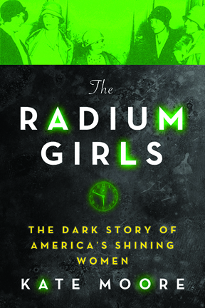
The legacy of the Radium Girls lives on through the ripples that their deaths created in labor law and our scientific understanding of the effects of radioactivity.
“Almost everything we know about radiation inside the human body, we owe to them,”
Radium Girls: The dark times of luminous watches
Jacopo Prisco, CNN 20th December 2017 A century ago, glow-in-the-dark watches were an irresistible novelty. The dials, covered in a special luminous paint, shone all the time and didn’t require charging in sunlight. It looked like magic.
One of the first factories to produce these watches opened in New Jersey in 1916. It hired about 70 women, the first of thousands to be employed in many such factories in the United States. It was a well-paid, glamorous job.
For the delicate task of applying the paint to the tiny dials, the women were instructed to point the brushes with their lips. But the paint made the watches glow because it contained radium, a radioactive element discovered less than 20 years earlier, its properties not yet fully understood. The women were ingesting it with nearly every brushstroke.
They became known as the “Radium Girls.”.
A miracle cure
Radium was discovered by Nobel laureate Marie Curie and her husband Pierre in 1898. It was quickly put to use as a cancer treatment.
“Because it was successful, it somehow became an all-powerful health tonic, taken in the same way as we take vitamins today — people were fascinated with its power,” said Kate Moore, author of “The Radium Girls,” in a phone interview………
A slow killer
When ingested, radium is particularly dangerous: “Chemically, it behaves very much like calcium,” said Jorgensen. “Since the body uses calcium to make bone, ingested radium is mistaken for calcium and gets incorporated into bone. So the major health risk of ingesting radium is radiation-induced bone necrosis and bone cancers. How soon they develop depends upon the dose, but at the very high doses that the Radium Girls were exposed to, just a few years.”
The luminous paint, which worked by converting the radiation into light through a fluorescent chemical, was one of the most successful radium-based products. By putting the brushes in their mouths, the Radium Girls were especially at risk — so why did they do it? “Because it was the easiest way to get a fine point on the brush, to paint on numbers as small as a single millimeter in width,” said Moore.
But the girls didn’t embrace this technique blindly. “The first thing they asked was (whether) the paint was harmful, but the managers said it was safe, which was the obvious answer for a manager of a company whose very existence depended on radium paint.”
Not all that glitters
When the luminous watches grew fashionable in the early 1920s, the world was already becoming aware of the risks of radioactivity. But radiation poisoning isn’t immediate, so years went by before any of the workers developed symptoms…….
Radium jaw
In the early 1920s, some of the Radium Girls started developing symptoms like fatigue and toothaches. The first death occurred in 1922, when 22-year-old Mollie Maggia died after reportedly enduring a year of pain. Although her death certificate erroneously stated that she died of syphilis, she was actually suffering from a condition called “radium jaw.” Her entire lower jawbone had become so brittle that her doctor removed it by simply lifting it out. “The radium was destroying the bone and literally drilling holes in the women’s jaws while they were still alive,” said Moore.
Yet it would take another two years before the company that owned the factory, the United States Radium Corporation, took any action at all, through an independent investigation commissioned mostly to investigate the declining business rather than the health of the workers.
In 1925 Grace Fryer, one of the workers from the original New Jersey plant, decided to sue, but she would spend two years searching for a lawyer willing to help her. She finally filed her case in 1927 along with four fellow workers, and made front-page news around the world.
The case, settled in the women’s favor in 1928, became a milestone of occupational hazard law. By this time, the dangers of radium were in full view, the lip-pointing technique was discontinued and the workers were being given protective gear. More women sued, and the radium companies appealed several times, but in 1939 the Supreme Court rejected the last appeal.
The survivors received compensation, and death certificates would start reporting the correct cause of death. The year before, the Food and Drug Administration banned the deceptive packaging of radium-based products. Radium paint itself was eventually phased out and has not been used in watches since 1968.
It’s hard to calculate how many women suffered health problems due to the ingestion of radium, but the certainly number in the thousands, according to Moore. Some of the effects would only be felt much later in life through various forms of cancer. With a half-life of 1,600 years, once the radium was inside the women’s bodies, it was there for good.
The legacy of the Radium Girls lives on through the ripples that their deaths created in labor law and our scientific understanding of the effects of radioactivity. “In the 1950s, during the Cold War, many agreed voluntarily to be studied by scientists, even with intrusive examinations because they had been exposed for prolonged periods of time,” said Moore.
Chris Busby published an answering to this paper. As soon as I am getting it, I will add it here below this paper.
By Bertrand R. Jordan – Unité Mixte de Recherche 7268 ADÉS, Aix-Marseille Université/Etablissement Français du Sang/Centre National de la Recherche Scientifique, Espace éthique méditerranéen, Hôpital d’Adultes la Timone, 13385 Marseille Cedex 05, France
ABSTRACT The explosion of atom bombs over the cities of Hiroshima and Nagasaki in August 1945 resulted in very high casualties, both immediate and delayed but also left a large number of survivors who had been exposed to radiation, at levels that could be fairly precisely ascertained. Extensive follow-up of a large cohort of survivors (120,000) and of their offspring (77,000) was initiated in 1947 and continues to this day. In essence, survivors having received 1 Gy irradiation ( 1000 mSV) have a significantly elevated rate of cancer (42% increase) but a limited decrease of longevity ( 1 year), while their offspring show no increased frequency of abnormalities and, so far, no detectable elevation of the mutation rate. Current acceptable exposure levels for the general population and for workers in the nuclear industry have largely been derived from these studies, which have been reported in more than 100 publications. Yet the general public, and indeed most scientists, are unaware of these data: it is widely believed that irradiated survivors suffered a very high cancer burden and dramatically shortened life span, and that their progeny were affected by elevated mutation rates and frequent abnormalities. In this article, I summarize the results and discuss possible reasons for this very striking discrepancy between the facts and general beliefs about this situation.
THEfirst (and only) two A-bombs used in war were deto-nated over Hiroshima and Nagasaki on August 6 and 9, 1945. Casualties were horrendous, approximately 100,000 in each city including deaths in the following days from severe burns and radiation. Although massive bombing of cities had already taken place with similar death tolls (e.g., Dresden, Hamburg, and Tokyo, the latter with 100,000 casualties on March 9, 1945), the devastation caused by a single bomb was unheard of and remains one of the most horrifying events in the past century. The people who had survived the explosions were soon designated as Hibakusha and were severely discrim-inated against in Japanese society, as (supposedly) carriers of (contagious?) radiation diseases and potential begetters of malformed offspring. While not reaching such extremes, the dominant present-day image of the aftermath of the Hiroshima/ Nagasaki bombings, in line with the general perception of radiation risk (Ropeik 2013; Perko 2014), is that it left the sites heavily contaminated, that the survivors suffered very serious health consequences, notably a very high rate of cancer and other debilitating diseases, and that offspring from these sur-vivors had a highly increased rate of genetic defects. In fact, the survivors have been the object of massive and careful long-term studies whose results to date do not support these conceptions and indicate, instead, measurable but limited det-rimental health effects in survivors, and no detectable genetic effects in their offspring. This Perspectives article does not provide any new data; rather, its aim is to summarize the results of the studies undertaken to date, which have been published in more than 100 papers (most of them in interna-tional journals), and to discuss why they seem to have had so little impact beyond specialized circles.
Bombings and Implementation of Cohort Studies
Characteristics of the bombs and the explosions
Figure 1 Number of solid cancers ob-served up to 1998 in the exposed group; the white portion indicates the excess cases associated with radiation (compar-ison with the unexposed group). Data are from Preston et al. (2007).
The device used at Hiroshima was based on enriched uranium and exploded at an altitude of 600 m with an estimated yield equivalent to 16 kilotons of high explosive. The bomb at Nagasaki was based on plutonium and exploded at 500 m with a yield of 21 kilotons. The major effect of both bombs was an extreme heat and pressure blast accompanied by a strong burst of gamma radiation and a more limited burst of neutrons. The heat blast set the (mostly wooden) buildings on fire in a radius of several kilometers and resulted in an extensive fire-storm centered on the explosion site (also called the hypocen-ter). People were exposed to the combined heat and radiation blasts, with little shielding from the buildings; most of those located within 1.5 km of the hypocenter were killed. The contribution of fallout from these explosions, which occurred mostly as “black rain” in the following days, is not precisely known: few measurements were taken due to scarcity of equipment, and investigations in the first months were per-formed by the US army and subsequently classified. It was probably limited: the bombs exploded at a significant altitude, the resulting firestorm carried the fission products into the high atmosphere, and the eventual fallout was spread over a large area. In addition, a strong typhoon occurred 2 weeks after the bombings and may have washed out much of the materiel. The major health effects (other than the heat blast and accompanying destruction) were almost certainly due to the gamma and neutron radiation from the blasts themselves, and these doses can be quite reliably estimated from the dis-tance to the hypocenter. Thus studies on the survivors can ascertain the health effects of a single, fairly well-defined dose of gamma radiation with a small component from neutrons.
The Atomic Bomb Casualty Commission and the Radiation Effects Research Foundation
Forbes 30th Oct 2017, Fukushima City is 50 miles northeast of the Fukushima-Daiichi Power Plant, so the radiation levels have been lower there than in the restricted areas, now reopening, that are closer to the plant. Hayama was unable to test monkeys in the most-contaminated areas, but even 50 miles from the plant,he has documented effects in monkeys that are associated with radiation.
He compared his findings to monkeys in the same area before 2011 and to a control population of monkeys in Shimokita Peninsula, 500 miles to the north. Hayama’s findings have been published in the peer-reviewed journal Scientific Reports, published by Nature.
Among his findings: Smaller Bodies — Japanese monkeys born in the path of fallout from the Fukushima meltdown weigh less for their height than monkeys born in the same area before the March, 2011 disaster, Hayama said. “We can see that the monkeys born from mothers who were exposed are showing low body weight in relation to their height, so they are smaller,” he said.
Smaller Heads And Brains — The exposed monkeys have smaller bodies overall, and their heads and
brains are smaller still. “We know from the example of Hiroshima and Nagasaki that embryos and fetuses exposed in utero resulted in low birth weight and also in microcephaly, where the brain failed to develop adequately and head size was small, so we are trying to confirm whether this also is happening with the monkeys in Fukushima,” Hayama said.
The WHOI research team also compared the radioactive contamination at the Marshall Islands to the contamination found today near Fukushima in Japan in the aftermath of the Dai-ichi Nuclear Power Plant disaster. “In contrast to Fukushima, where cesium is the most abundant radionuclide of concern, in these atolls, the focus should be on plutonium, given its significantly high levels,” said WHOI radiochemist Ken Buesseler.
Scientists have found lingering radioactivity in the lagoons of remote Marshall Island atolls in the Pacific Ocean where the United States conducted 66 nuclear weapons tests in the 1940s and 1950s.
Radioactivity levels at Bikini and Enewetak Atolls were extensively studied in the decades after the testing ended, but there has been relatively little work conducted there recently. A team of scientists from Woods Hole Oceanographic Institution (WHOI) reported that levels of radioactive cesium and plutonium have decreased since the 1970s, but these elements continue to be released into the Pacific Ocean from seafloor sediments and lagoon waters.
The levels of plutonium are 100 or more times higher in lagoon waters compared to the surrounding Pacific Ocean and about two times higher for a radioactive form of cesium. Despite these enrichments, they do not exceed U.S. and international water quality standards set to protect human health, the scientists reported Oct. 30, 2017, in the journal Science of the Total Environment.
To determine the source of these radionuclides in lagoon waters, the WHOI scientists measured the amounts and flow of radioactive material entering the ocean from groundwater seeping from the islands. They found that groundwater was a relatively low source of radioactivity.
In particular, they found that radioactive groundwater was not leaking much from beneath one suspected potential source: the Runit Dome on the island of Runit—a massive 350-foot-wide concrete lid that covers 111,000 cubic yards of radioactive soil and debris that were bulldozed into a bomb crater and sealed over. It was constructed in the late 1970s by the U.S. government to contain contaminated waste from the nuclear tests. The bottom of the Runit Dome is not lined and below sea level, so scientists and others have been concerned that tidal action could move water through the buried radioactive material and bring it out to sea.
“The foundations of these island atolls are ancient coral reefs that have the porosity of Swiss cheese, so groundwater and any mobilized radioactive elements can percolate through them quite easily,” said WHOI geochemist Matt Charette. Though that does not seem to be happening now, the scientists advise that the Runit Dome area should be continuously monitored as sea level rises and the dome deteriorates.
Using isotopes of plutonium that act like a fingerprint to pinpoint sources, the WHOI scientists found that the seafloor sediments around Runit Island seem to be contributing about half of the plutonium to the lagoon. “Additional studies examining how radioactive plutonium moves through the environment would help elucidate why this small area is such a large source of radioactivity,” Buesseler said.
The WHOI scientists who conducted the study and wrote the report included Ken Buesseler, Matthew Charette, Steven Pike, Paul Henderson, and Lauren Kipp. They sailed to the islands aboard the research vessel Alucia on an expedition funded by the Dalio Explore Fund.
The team collected sediments from the lagoon with poster tube-sized collectors that were inserted by divers into the seafloor’s sediments, filled with mud, capped. Back in WHOI laboratories, the cores were sliced into layers and analyzed to reveal a buried record of local fallout from the nuclear tests. The scientists also collected and analyzed samples of lagoon waters .
On the islands, they collected groundwater samples from cisterns, wells, beaches, and other sites. They analyzed these samples for the levels of radioactive cesium and plutonium from weapons tests. For the first time on these islands, the scientists also measured isotopes of radium, a naturally occurring radioactive “tracer” that give scientists key information to determine how much and how fast groundwater flows from land into the ocean.
The WHOI research team also compared the radioactive contamination at the Marshall Islands to the contamination found today near Fukushima in Japan in the aftermath of the Dai-ichi Nuclear Power Plant disaster. “In contrast to Fukushima, where cesium is the most abundant radionuclide of concern, in these atolls, the focus should be on plutonium, given its significantly high levels,” said WHOI radiochemist Ken Buesseler.
The U.S. conducted 66 nuclear weapons tests between 1946 and 1958 at Bikini and Enewetak Atolls, each a ring of low-lying reef islands that surrounds a larger lagoon. Bikini has 26 islands; Enewetak had 42 islands, but three were bombed out of existence. They became known as the western part of the “U.S. Pacific Proving Grounds.”
Bikini and Enewetak are among 29 atolls that make up the Republic of the Marshall Islands, located in the equatorial Pacific, about 2,500 miles west of Hawaii. The collective land area of the thousands of small islands is equivalent to the area of Washington, D.C. but they are spread across an ocean area that exceeds the size of Alaska.
The work holds particular significance to the atolls’ indigenous populations which were evacuated before the tests and thus far have only been allowed to return to one small island in the Enewtak Atoll.
This research was funded by the Dalio Foundation and the Dalio Explore Fund.
The Woods Hole Oceanographic Institution is a private, non-profit organization on Cape Cod, Mass., dedicated to marine research, engineering, and higher education. Established in 1930 on a recommendation from the National Academy of Sciences, its primary mission is to understand the ocean and its interaction with the Earth as a whole, and to communicate a basic understanding of the ocean’s role in the changing global environment. For more information, please visit www.whoi.edu.