Link above is to a PDF, 47 page, readable report. Very well done. stock out
Timothy Mousseau to lead research into radiation in Chernobyl dogs
South Carolina researcher wants to learn about radiation by researching stray dogs near Chernobyl https://www.postandcourier.com/features/south-carolina-researcher-wants-to-learn-about-radiation-by-researching/article_eee33baa-5454-11e8-89c1-17da43576a65.html By Mary Katherine Wildeman mkwildeman@postandcourier.com May 14, 2018
Japan’s planned Olympics torch relay route found to have high levels of radiation
the proposed torch route samples had the highest mean radioactivity due to their close proximity to the plant. Based on the measurement, we estimated qualitatively that the radiation exposure of people living near the Azuma Sports Park area was 20.7 times higher than that of people living in Tokyo. The main tourist and proposed torch routes had radiation exposure of 24.6 and 60.6 times higher, respectively, than in Tokyo.
our results showed that the highest radioactivity level was at the proposed torch route, which is located to the northeast of the plant.
Olympic officials should consider using the results of this project to decide whether the radioactivity level at the proposed torch route and the Olympic venues are within acceptable level.

Measuring Radioactivity in Soil and Dust Samples from Japan Fukushima IQP Final Report. An Interactive Qualifying Project Report Submitted to the Faculty of the Worcester Polytechnic Instituteby Thang Pham Steven Franca Son Nguyen
Abstract
A radioactivity map of Cesium-137, a radioactive material emitted from the Fukushima Dai-ichi Nuclear Power Plant, was created by examining 30 soil and dust samples originating from Japan. A Sodium Iodide (NaI) detector measured gamma rays emitted from the samples. Qualitative evaluation of human exposure to radio-cesium in five sets of locations in Japan shows the radioactivity of tested samples. The Fukushima section of one of the Olympic proposed torch route has a 60 times higher activity of Cesium-137 than the activity of the samples from Tokyo.
Acknowledgements…….
Executive Summary
Overview
On March 11th, 2011, a magnitude 9.0 earthquake hit the Fukushima Prefecture of Japan (AESJ, 2015, p.19). This resulted in major structural damage to the surrounding area. Following the earthquake, a 13-meter tsunami hit the Fukushima Dai-ichi Nuclear Power Plant and disabled the backup the diesel backup power generator and the cooling water pumps needed to keep the Power Plant running in the event of a disaster. Since power restoration was unsuccessful and there was major structural damage caused to the Units by the tsunami, there were high concentrations of nuclear particles released into the air.
The amount of radioactive materials emitted from the Power Plant raised health and environmental concerns to the people living in the areas around the Fukushima Dai-ichi Nuclear Power Plant. In fact, several studies conducted after the accident determined the activity of radioactive isotopes in the environments. These studies around Fukushima provided excellent understanding of the activity of radioactive isotopes in the environment and the health effects on humans.
With the upcoming XXXII Olympiad in 2020 hosted by Japan, it is necessary to look into the radioactivity of Olympic venues as well as tourist attractions in the host cities. Previous studies pose a major gap because they focused on regions that are closer to the Fukushima Daiichi Nuclear Power Plant, where the highest concentration of radioactive fallout is located. On the other hand, the radioactivity in other locations in Japan is relatively less studied. Since thousands of athletes and millions of visitors are travelling to Japan for the Olympics, there has been widespread concern from the international community about radiation exposure. Therefore, it is important to investigate the extent of radioactive fallout from the Fukushima Dai-ichi incident.
Project goals and Objectives
The goals of the project were to measure the radioactivity of Cesium-137 in soil and dust samples from Japan and to generate a map that illustrates the activity of Cesium-137 in five locations in Japan.Understanding the radioactivity in Japan soil would help in giving an estimation of human exposure to radioactivity in the studied areas. The primary objectives of this project were to:
- Perform soil sample preparation for measurement, including categorizing, drying and sub-sampling
- Measure the activity of Cesium-137 in the samples and analyze the radioactivity at different locations in Japan.
- Generate a map to illustrate the radioactivity level in Japan soil.
- Estimate qualitatively human exposure to radioactivity for people living in the studied areas.
Development of methodology
The employment of various study methods and experiment designs helped complete the objectives. Through literature review and discussion with advisors and lab instructors, we were able to develop main steps and setting for our study.
Primarily, we performed preparation for 87 soil and dust samples collected in Japan and transferred to Worcester Polytechnic Institute (WPI). The samples were classified into five categories based on their original location: Tokyo samples, Azuma Sports Park samples, proposed torch route samples, tourist route samples and non-Olympic samples. All the samples were dried and sub-sampled so that their physical conditions were consistent throughout the testing phase. From the 87 samples that are dried and sub-sampled, we eventually further narrowed to 30 samples for radioactivity measurement.
Previous studies on radioactive nuclides accumulation in soil and dust samples from Japan identified that Cesium-137 had the highest concentration among radionuclides in Japan soil (Saito et al., 2015). Therefore, we set our goal to measure the activity of Cesium-137 in the samples from Japan. A thallium-activated sodium iodine detector, or NaI (TI) detector, measured the concentration of Cesium-137 in the soil samples. The calibrating sources used for Cesium137 measurement were Cesium-137, Sodium-22 and Manganese-54. The experiment reported the radioactivity of the background and the radioactivity of Cesium-137 in the samples above the background. Measurements were made in triplicate to provide counting variability statistics.
At the beginning of the project, samples were selected randomly for measurement. Our focus then shifted, as we looked deeper into the four main areas that will be important to the XXXII Olympiad in 2020. These Olympic areas included Azuma Sports Park, tourist attractions in Tokyo, a proposed torch route, and official designated tourist routes. Time constraint and large amount of samples, limited testing which resulted in results on only a limited part of each sample set.
After finishing measuring, the radioactivity of the soil and dust samples helped generate a map that shows the activity of Cesium-137 in Japan soil. Since there was a large variation in the measured activity between samples at different locations, we decided to analyze the results based on the samples’ original locations.
Our results were cross-referenced with information and results gathered from literature reviews. Understanding the activity of Cesium-137 in Japan soil, we were able to give a qualitative estimation of human exposure.
Results and Analysis
By the end of the project, we were able to measure the activity of Cesium-137 in 30 soil and dust samples. The results showed a great variation in radioactivity between samples from different locations. The activity of Cesium-137 in the 30 samples ranged from 38.6 Bq/kg to 28041.5 Bq/kg with the mean radioactivity of 4903.3 Bq/kg (standard deviation, σ= 6611.0 Bq/kg).
Among the five categories of locations, the Tokyo sample set had the lowest radioactivity mean of 117.6 Bq/kg. The following locations are ordered by increasing mean of radioactivity: Azuma Sports Park samples (2703.9 Bq/kg), main tourist routes samples (3206.0 Bq/kg) and proposed torch route samples (7896.0 Bq/kg). Only one of the non-Olympic samples, which originates from the Entrance to Fukushima Dai-ichi waste mounds, had a radioactivity of 14119.8 Bq/kg. Due to this outlier, this sample set was omitted from the other sets.
The measured results showed that the Tokyo sample set had the lowest radioactivity level, which was understandable based on its significant distance from the Fukushima Nuclear Power Plant. On the contrary, the proposed torch route samples had the highest mean radioactivity due to their close proximity to the plant. Based on the measurement, we estimated qualitatively that the radiation exposure of people living near the Azuma Sports Park area was 20.7 times higher than that of people living in Tokyo. The main tourist and proposed torch routes had radiation exposure of 24.6 and 60.6 times higher, respectively, than in Tokyo.
The results in this study were in agreement with published results qualitatively. Saito et al. reported a much higher deposition of Cesium-137 in the area to the northeast of the Fukushima Nuclear Power Plant and the further away from the plant, the lower the deposition of Cesium-137 (2015). Accordingly, our results showed that the highest radioactivity level was at the proposed torch route, which is located to the northeast of the plant. The further away from the plant for example, in Tokyo and the Azuma Sports Park, the lower the activity of Cesium137. However, because of the differences in sampling techniques and reported unit of radioactivity, there could not be an established direct comparison.
Recommendations
Given the aforementioned results, there is significant evidence through our testing, that there are still high amounts of radiation in areas close to the Fukushima Prefecture. Past and current tests found that high amounts of radiation remain at the origin of the disaster. The results showed a significantly higher amount of radioactive materials in the proposed torch route compared to those in Tokyo.
Given the results from testing, we confidently make the following recommendations:
- Further study on the activity of Cesium-137 on the proposed torch route is necessary to understand how much radioactive materials remain.
- A more detailed study with more soil and dust samples can give a more accurate map of specific areas of interest.
- Olympic officials should consider using the results of this project to decide whether the radioactivity level at the proposed torch route and the Olympic venues are within acceptable level. 4. Qualified health professionals should use this data to make connections about short and long-term effects on health.
Authorship……….
Table of Contents……. 47 pages Very well supplied with references, diagrams, maps, … https://web.wpi.edu/Pubs/E-project/Available/E-project-031418-011249/unrestricted/Fukushima_IQP_Final_Report.pdf
There’s money in monitoring ionising radiation

 Radiation Detection, Monitoring & Safety Market Worth 2.26 Billion USD by 2022 PUNE, India, May 8, 2018 /PRNewswire/ —
According to a new market research report “Radiation Detection, Monitoring, & Safety Market by Product (Detection & Monitoring, Safety), Composition (Gas-filled detectors, Scintillator, Solid-state detector), Application (Healthcare, Homeland Security & Defence, Industrial) – Global Forecast to 2022“, published by MarketsandMarkets™, the global market is expected to reach USD 2.26 Billion by 2022 from USD 1.71 Billion in 2017, at a CAGR of 5.7% during the forecast period (2017-2022).
The key factors propelling the growth of Radiation Detection, Monitoring and Safety Market are growing security threats, growing prevalence of cancer worldwide, increasing safety awareness among people working in radiation-prone environments, growing safety concerns post the Fukushima disaster, growing security budgets of global sporting events, growth in the number of PET/CT scans, increasing usage of nuclear medicine and radiation therapy for diagnosis and treatment, and use of drones for radiation monitoring.
….The healthcare segment dominated the market on the basis of applications in 2017
The Radiation Detection, Monitoring and Safety Market is segmented on the basis of applications into healthcare, homeland security and defense, industrial applications, nuclear power plants, and other applications (environmental monitoring and academic research). In 2017, the healthcare segment accounted for the largest share of the global market. Factors such as the growth in the number of PET/CT scans and increasing usage of nuclear medicine and radiation therapy for diagnosis and treatment, increasing research activities, and growing incidence of cancer are driving the growth of this segment.
North America held the largest share of the market in 2017
In 2017, North America accounted for the largest share of the Radiation Detection, Monitoring and Safety Market, followed by Europe. Factors such as favorable government initiatives, increasing number of nuclear power plants, rising prevalence of cancer, and increasing awareness of radiation safety are contributing to the large share of this geographical segment………https://www.prnewswire.com/news-releases/radiation-detection-monitoring–safety-market-worth-226-billion-usd-by-2022-682063101.html
U.S. Department of Energy sending helicopters to measure radiation levels
UF flyovers part of DOE radiation study http://www.gainesville.com/news/20180508/uf-flyovers-part-of-doe-radiation-study
The flyovers are part of a research project measuring baseline levels of radiation in the environment.
The U.S. Department of Energy’s National Nuclear Security Administration says the public shouldn’t be alarmed by a low-flying helicopter near the University of Florida.
It’s a part of a routine procedure, officials say.
According to a Department of Energy press release, the public might see a twin-engine Bell 412 helicopter flying at about 150 feet or higher around UF and in Gainesville until early Wednesday evening.
The helicopter is operated by the Remote Sensing Laboratory Aerial Measuring System from Joint Base Andrews in Prince George’s County, Maryland, and is equipped with radiation-sensing technology.
The manned helicopter will fly in a grid pattern over the area at about 80 miles per hour, the release says.
The flyovers are part of a research project measuring baseline levels of radiation in the environment.
How safe is cellphone’s electromagnetic radiation?
 Why does this article not mention the difference in vulnerability between children and adults?
What is mobile phone radiation and how safe is it? ABC Science,By science reporter Belinda Smith
“…. What is electromagnetic radiation?

We are surrounded by all sorts of different types of electromagnetic radiation every day: your eyes pick up visible light, your bag is scanned by X-rays at airport security, microwaves heat your lunch and too much ultraviolet light gives you sunburn.
At its essence, electromagnetic radiation is energy comprising an electric field and magnetic field, which travel together, but perpendicularly, in waves.
Sometimes the length of these waves (or wavelength) is very short — a few nanometres for X-rays — while others are much longer — a few centimetres up to kilometres.
It’s these long wavelengths, called radio waves, that are the electromagnetic radiation of choice for mobile phones and base stations.
Unlike shorter wavelengths, such as visible light, radio waves can pass through walls. The longer the wavelength, the better it can penetrate solid stuff.
Another term you might see is frequency, which is the number of times a wave makes a full oscillation each second.
Frequency and wavelength are closely related. Wavelength is the speed of light divided by the frequency, so long wavelengths also have low frequency.
What are ionising and non-ionising radiation?

The radio frequency end of the electromagnetic spectrum is home to what’s known as “non-ionising radiation”, said Rodney Croft, from the University of Wollongong and director of the Australian Centre of Electromagnetic Bioeffects Research.
It’s the high-frequency, short wavelength radiation, such as X-rays, that can tinker with your DNA and are linked to cancer.
These waves are small enough and carry enough energy to knock electrons off atoms, ionising them.
Radio frequency used in mobile communications simply doesn’t have the energy to do that. But that’s not to say it doesn’t exert any effects on the matter it travels through.
“It’s an oscillating wave, which swings between positive and negative,” Professor Croft said
“If you have a bunch of molecules rotating, that causes friction, and energy is given off as heat. It’s how a microwave oven works.”
Does anyone regulate radio frequency limits?
In Australia, mobile phone and base station exposure limits are set by the Australian Radiation Protection and Nuclear Safety Agency (ARPANSA) and regulated by the Australian Communications and Media Authority.
The ARPANSA standard looks at how much energy a user absorbs from a mobile phone over time.
The maximum limit is currently 2 watts per kilogram of tissue. Phone manufacturers usually specify their maximum absorption rate in their manual.
You can find how much radio frequency is emitted by base stations at the Radio Frequency National Site Archive.
According to ARPANSA, it’s “typically hundreds of times below the [ARPANSA] limits”.
What are the effects of mobile radio frequency on tissues?
So are we microwaving our head whenever we answer the phone? A tiny bit, but not enough to be worried about, Professor Croft said.
He and his team found mobile phone radiation exposure increases the temperature of the outer grey, wrinkled layer of the brain called the cortex, but it’s only “maybe about 0.1 degree, which is very small compared to the temperature variation the body normally has to contend with”, he said.
“We do find that we get a slight change to thermoregulation, so the body, even with that small change, is sending a bit more blood out to the periphery to cool it, so your body doesn’t end up warming up.”
……...What about cancer in rodents?
Mobile phones are classed as “possibly carcinogenic” by the International Agency for Research on Cancer, putting them in the same category as aloe vera, bracken fern and Asian pickled vegetables.
And while radio frequency is non-ionising radiation — remember, it can’t knock electrons off atoms, fiddle with genetic material and trigger tumour growth like ionising radiation can — studies still investigate possible links.
Research published in February this year by the US National Toxicology Program found tumours grew in the nerves around the heart of male rats if they were bathed in extremely high levels of mobile radiation.
But, Professor Croft said, “there were so many difficulties with that study.
………Risks and benefits
Despite research showing no link between safe levels of radio frequency and cancer, telecommunications companies and other organisations do offer suggestions if you want to reduce exposure.
The obvious action, Dr Halgamuge said, is to limit mobile phone use: “You have no control over base stations, because that radiation is around you all the time, but you do have control over your mobile phone.”
The ARPANSA also recommends using hands-free or texting instead of calling, “but none of those things are actually based on any health effects”, Professor Croft said.
………So: does radio frequency have any effect on human tissue, apart from heating it a fraction of a degree?
That question is still open, Professor Wood said.
“Even though some of the evidence [that radio frequency causes damage] on the face of it looks quite compelling, there’s still a question of consistency…….http://www.abc.net.au/news/science/2018-04-30/mobile-phone-radiation-health-explainer-biophysics/9702630
New Study Shows Full Extent of Radiation Damage to Hiroshima Victims
https://www.popularmechanics.com/military/weapons/a20113160/new-study-shows-full-extent-of-radiation-damage-to-hiroshima-victims/ A study decades in the making shows victims may have absorbed double a deadly dose. By David Grossman
A weapon drastically different than any other ever used in war, the atomic bomb in Hiroshima instantly killed over 100,000 people and left thousands more dealing with radiation fallout. By the end of 1945, it is estimated that 160,000 people had been killed directly from the bombing. Several historians have argued that while the bombs effectively ended World War II, their unprecedented destructive capabilities started the next global conflict, the Cold War, at the exact same time.
Attempting to measure the damage done to Hiroshima by the atomic bomb overwhelmed science for decades. There were simply no computers or radiation-measuring devices capable of understanding the damage. Personal stories, like those of the survivors describe in John Hershey’s Hiroshima and art works of survivors, took hold as the dominant narratives.
But that didn’t mean scientists weren’t trying. When the Atomic Bomb Casualty Commission (ABCC) formed in 1947, the agency quickly realized it would need long term study to understand what had happened. Japanese scientists like E. T. Arakawa and Takenobu Higashimura were releasing studies about the effects of the bombings by the early 1960s.
In 1973, Brazilian physicist Sérgio Mascarenhas was trying to date archaeological items in his home country based on radiation absorption. Radiation occurs naturally in sand through elements like thorium, and techniques like radiocarbon dating use similar principles.
However, Mascarenhas realized that this method might have applications beyond archaeological items. He flew to Hiroshima and, with help from the Institute of Nuclear Medicine in Hiroshima, was able to obtain a jawbone from a bombing victim’s body. While he gained some understanding of what the victim’s body had endured, technical issues stood in his way. He was unable to separate background radiation levels from the bomb blast radiation.
Flash forward four decades later and Angela Kinoshita of Universidade do Sagrado Coração in São Paulo State has reexamined the jawbone using modern technology. Kinoshita’s team was able to determine that the jawbone absorbed 9.46 grays of radiation. A mere 5 grays can be fatal. That number lines up with measurements taken of bricks and other inorganic objects measured at the time. The work is published in PLOS ONE.
Beyond gaining a better understanding of what happened to the victims of Hiroshima, who ranged from prisoners of war to soldiers to civilians, the study offers insight into what might happen if a nuclear weapon was ever used again.
“Imagine someone in New York planting an ordinary bomb with a small amount of radioactive material stuck to the explosive. Techniques like this can help identify who has been exposed to radioactive fallout and needs treatment,” says study co-author Oswaldo Baffa of the University of São Paulo in a press statement. Source: Discover
In new technique, scientists calculate radiation dose in bone from victim of Hiroshima bombing
Scientists calculate radiation dose in bone from victim of Hiroshima bombing https://www.eurekalert.org/pub_releases/2018-04/fda-scr042718.php
In an article published in PLOS ONE, Brazilian researchers describe the first retrospective dosimetric study by electron spin resonance spectroscopy using human tissue from nuclear attack victimsFUNDAÇÃO DE AMPARO À PESQUISA DO ESTADO DE SÃO PAULO
The bombing of the Japanese cities Hiroshima and Nagasaki by the United States in 1945 was the first and only use of nuclear weapons against civilian targets. A series of studies began in its aftermath to measure the impact of the fallout, in terms of both the radiation dose to which the victims were exposed and the effects of this exposure on DNA and health in general.
Continuing research that started in the 1980s under the leadership of physicist Sérgio Mascarenhas, Full Professor at the University of São Paulo (USP), Brazilian scientists have published an article in the journal PLOS ONE describing a method of precise measurement of the radiation dose absorbed by the bones of victims of the nuclear bombs dropped on Japan.
The investigation was conducted during the postdoctoral research of Angela Kinoshita, currently a professor at Universidade do Sagrado Coração in Bauru, São Paulo State. Her supervisor was then Oswaldo Baffa, Full Professor at the University of São Paulo’s Ribeirão Preto School of Philosophy, Science & Letters (FFCLRP-USP).
“We used a technique known as electron spin resonance spectroscopy to perform retrospective dosimetry. Currently, there’s renewed interest in this kind of methodology due to the risk of terrorist attacks in countries like the United States,” Baffa said.
“Imagine someone in New York planting an ordinary bomb with a small amount of radioactive material stuck to the explosive. Techniques like this can help identify who has been exposed to radioactive fallout and needs treatment.”
As Kinoshita explained, the study is unique insofar as it used samples of human tissue from victims of the bomb dropped on Hiroshima.
“There were serious doubts about the feasibility of using this methodology to determine the radiation dose deposited in these samples, because of the processes involved in the episode,” she said. “The results confirm its feasibility and open up various possibilities for future research that may clarify details of the nuclear attack.”
The equipment used in the investigation was purchased during a project coordinated by Baffa and supported by the São Paulo Research Foundation – FAPESP.
Origins
In the 1970s, when he was teaching at the University of São Paulo’s São Carlos Physics Institute (IFSC-USP), Mascarenhas discovered that X-ray and gamma-ray irradiation made human bones weakly magnetic. The phenomenon, known as paramagnetism, occurs because the hydroxyapatite (crystalline calcium phosphate) in the mineral portion of bone tissue absorbs carbon dioxide ions, and when the sample is irradiated, the CO2 loses electrons and becomes CO2-. This free radical serves as a marker of the radiation dose received by the material.
“I discovered that we could use this property to perform radiation dosimetry and began using the method in archeological dating,” Mascarenhas recalled.
His aim at the time was to calculate the age of bones found in sambaquis (middens created by Brazil’s original inhabitants as mounds of shellfish debris, skeletons of prehistoric animals, human bones, stone or bone utensils, and other refuse) based on the natural radiation absorbed over centuries via contact with elements such as thorium that are present in the sand on the seashore.
On the strength of this research, he was invited to teach at Harvard University in the United States. Before leaving for the US, however, he decided to go to Japan to try to obtain samples of bones from victims of the nuclear bombs and test his method on them.
“They gave me a jawbone, and I decided to measure the radiation right there, at Hiroshima University,” he said. “I needed to prove experimentally that my discovery was genuine.”
Mascarenhas succeeded in demonstrating that a dosimetric signal could be obtained from the sample even though the technology was still rudimentary and there were no computers to help process the results. The research was presented at the American Physical Society’s annual March Meeting, where it made a strong impression. Mascarenhas brought the samples to Brazil, where they remain.
“There have been major improvements in the instrumentation to make it more sensitive in the last 40 years,” Baffa said. “Now, you see digitally processed data in tables and graphs on the computer screen. Basic physics has also evolved to the extent that you can simulate and manipulate the signal from the sample using computational techniques.”
Thanks to these advances, he added, in the new study, it was possible to separate the signal corresponding to the radiation dose absorbed during the nuclear attack from the so-called background signal, a kind of noise scientists suspect may have resulted from superheating of the material during the explosion.
“The background signal is a broad line that may be produced by various different things and lacks a specific signature,” Baffa said. “The dosimetric signal is spectral. Each free radical resonates at a certain point on the spectrum when exposed to a magnetic field.”
Methodology
To make the measurements, the researchers removed millimeter-scale pieces of the jawbone used in the previous study. The samples were again irradiated in the laboratory using a technique called the additive dose method.
“We added radiation to the material and measured the rise in the dosimetric signal,” Baffa explained. “We then constructed a curve and extrapolated from that the initial dose, when the signal was presumably zero. This calibration method enabled us to measure different samples, as each bone and each part of the same bone has a different sensitivity to radiation, depending on its composition.”
Thanks to this combination of techniques, they were able to measure a dose of approximately 9.46 grays (Gy), which is high in Baffa’s view. “About half that dose, or 5 Gy, is fatal if the entire body is exposed to it,” he said.
The value was comparable with the doses obtained by other techniques applied to non-biological samples, such as measurement of the luminescence of quartz grains present in brick and roof tile fragments found at the bomb sites. According to the authors, it was also close to the results of biological measurement techniques applied in long-term studies using alterations in survivors’ DNA as a parameter.
“The measurement we obtained in this latest study is more reliable and up to date than the preliminary finding, but I’m currently evaluating a methodology that’s about a thousand times more sensitive than spin resonance. We’ll have news in a few months,” Mascarenhas predicted.
About São Paulo Research Foundation (FAPESP)
The São Paulo Research Foundation (FAPESP) is a public institution with the mission of supporting scientific research in all fields of knowledge by awarding scholarships, fellowships and grants to investigators linked with higher education and research institutions in the State of São Paulo, Brazil. FAPESP is aware that the very best research can only be done by working with the best researchers internationally. Therefore, it has established partnerships with funding agencies, higher education, private companies, and research organizations in other countries known for the quality of their research and has been encouraging scientists funded by its grants to further develop their international collaboration. For more information: http://www.fapesp.br/en.
ULTRAVIOLET RADIATION FROM LOW-MASS STARS COULD RENDER PLANETS UNINHABITABLE
 Astrobiology Magazine, By Amanda Doyle – Apr 19, 2018
Low-mass stars are currently the most promising targets when searching for potentially habitable planets, but new research has revealed that some of these stars produce significant amounts of ultraviolet (UV) radiation throughout their lifetimes. Such radiation could hinder the development of life on any orbiting planets.
M-dwarfs are stars that are cooler and less massive than stars like our Sun, and are the most common type of star in the Galaxy, meaning that it is vital that we better understand them and the influence they have on their planets.
Detecting terrestrial planets in the habitable zone – the region where liquid water can exist on a planet’s surface – when they pass in front of, or transit, Sun-like stars is difficult. This is partly because we only see a small dip in the light as the planet crosses the star, and also partly because their orbits are long enough that we have to wait several years to observe multiple transits. However, because M-dwarfs are smaller and cooler, the planets in their habitable zone are much closer to their star, resulting in larger and more frequent drops in light, making them easier to detect.
This makes M-dwarfs ideal candidates when searching for potentially habitable planets, which has led to habitable zone terrestrial planets being discovered around M-dwarfs including Proxima Centauri, TRAPPIST-1and Ross 128.
Ultraviolet levels over time
A paper by astrophysicists Adam Schneider and Evgenya Shkolnik from Arizona State University, recently published in The Astronomical Journal,has revealed that the hottest and most massive M-dwarfs, referred to as ‘early type’, emit different amounts of UV radiation over their lifetime compared to the less massive and cooler ‘mid-’ and ‘late-type’ M-dwarfs. The paper used observations from NASA’s Galaxy Evolution Explorer (GALEX) spacecraft to study several populations of M-dwarfs in ultraviolet light.
M-dwarfs are known to emit higher levels of potentially harmful UV radiation than stars like our Sun. UV radiation can erode planetary atmospheres and have a detrimental effect on biology. It can also affect the abundances of molecules in planetary atmospheres, including carbon dioxide, oxygen and ozone……….
Harmful to life
The results suggest that lower-mass M-dwarfs have persistent UV radiation, which could rule out the possibility of life on any orbiting planets, including those around TRAPPIST-1 and Proxima Centauri………https://www.astrobio.net/alien-life/ultraviolet-radiation-from-low-mass-stars-could-render-planets-uninhabitable/
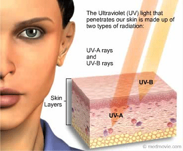
The Mechanism of DNA Damage by UV Radiation
 News Medical Life Sciences,
Solar ultraviolet radiation (UV) exposure triggers DNA damage, a preliminary step in the process of carcinogenesis.
The stability of DNA is extremely important for the proper functioning of all cellular processes. Exposure to UV radiation alters the structure of DNA, affecting the physiological processes of all living systems ranging from bacteria to humans.
Ultraviolet Radiation
Natural sunlight stimulates the production of vitamin D, an important nutrient for the formation of healthy bones. However, sunlight is also a major source of UV radiation. Individuals who get excessive UV exposure are at a great risk of developing skin cancers. There are three types of UV rays: UVA, UVB and UVC.
- UVC rays (100-280 nm) are the most energetic and damaging of the three rays. Fortunately, UVC is absorbed by the ozone layer before reaching the earth’s surface.
- UVA rays (315-400 nm) possess the lowest energy and is able to penetrate deep into the skin. Prolonged exposure has been linked to ageing and wrinkling of the skin. UVA is also the main cause of melanomas.
- UVB rays (280-315 nm) possess higher energy than UVA rays and affect the outer layer of the skin leading to sunburns and tans. Basal cell carcinoma and squamous cell carcinoma are caused by UVB radiation.
DNA Damage by UV Radiation
DNA is composed of two complementary strands that are wound into a double helix. The hereditary message is chemically coded and made up of the four nucleotides adenine (A), thymine (T), guanine (G) and cytosine (C). UVB light interferes directly with the bonding between the nucleotides in the DNA. ……….
https://www.news-medical.net/life-sciences/The-Mechanism-of-DNA-Damage-by-UV-Radiation.aspx
Radiation contamination of 45 Hanford nuclear workers
42 Hanford workers contaminated with radiation, Seattle Times, March 24, 2018 The final results of worker tests after a December spread of contamination found that 11 Hanford workers had inhaled or ingested radioactive particles from demolition of the nuclear reservation’s Plutonium Finishing Plant. By Annette Cary Tri-City Herald ioactive contamination from demolition of the nuclear reservation’s Plutonium Finishing Plant.
Increased UV radiation brings increased skin cancer risk
Going to extremes: UV radiation is on the way up, https://www.smh.com.au/environment/weather/going-to-extremes-uv-radiation-is-on-the-way-up-20180308-p4z3cp.htmlSMH, Nigel Gladstone
The combination of a thinning ozone layer and farming practices in India may add up to more days of extreme ultraviolet radiation across Australia.
A Sun-Herald analysis of daily UV index readings since 1997 in Sydney, Melbourne and Brisbane found the number of days when ultraviolet radiation reached or passed extreme levels had risen slightly.
The amount of UV that hits Australia is influenced by fluctuations in cloud cover, ozone levels and the solar cycle.
In Sydney, four of the 10 highest UV index days since 1996 have been recorded since December 2016. While the ozone layer is recovering over the poles, it is thinning in mid-latitudes from Russia to the Southern Ocean below Australia, a study published last month in the journal Atmospheric Chemistry and Physics found.
“Decreases in ozone are less than we saw at the poles before the Montreal Protocol was enacted [in 1987], but UV radiation is more intense in these regions and more people live there,” said report co-author Joanna Haigh, from Imperial College London.
The weather bureau studied UV radiation in Australia between 1959 and 2009 and found an annual increase of 2 to 6 per cent since the 1990s, above a 1970-80 baseline. The bureau found these changes were related to ozone depletion.
Associate Professor Clare Murphy, from the school of chemistry at Wollongong University, said ozone trends were not fully understood.
“The largest factor involved in mid-latitude ozone depletion is the nitrogen cycle, which operates by nitrous oxide turning into reactive nitrogen in the stratosphere,” Dr Murphy said.
Nitrogen fertiliser is converted into nitrous oxide by soil microbes, creating a stable greenhouse gas that can reach the stratosphere, where the ozone layer protects the earth from most of the sun’s UV radiation,” she said. “However, once in the stratosphere, nitrous oxide is broken down by high energy radiation from the sun to become reactive nitrogen, which can deplete ozone.”
Dr Murphy said that last century, concerns about ozone depletion centred on “chlorine chemistry” (CFCs) because of the massive hole over the poles. “Now it’s nitrous oxide, which almost stopped the Concord from flying because they were worried about reactive nitrogen in the stratosphere.”
Nitrous oxide damage to ozone is ubiquitous, whereas damage from CFCs creates a hole during extreme weather years over the Antarctic, Dr Murphy said.
Nitrous oxide was identified as the most damaging substance to the ozone layer in the 21st century by a 2009 study published in Science. That study also suggested one of the best ways to address the problem was to give insurance to Indian farmers.
“In India, particularly, they’re putting in 10 times more nitrogen fertiliser on their crops than they need to because if a crop fails they may starve,” Dr Murphy said. “Insurance could pick up the loss.”
Robin Schofield, director of Melbourne University’s environmental science hub, said UV in Australia should be trending downwards because factors such as surface ozone, which is contained in smog, is on the rise and there is evidence of a recovery of stratospheric ozone.
The UV Index and skin cancer
The UV index relates to the intensity of sunburn-producing UV radiation. Sun protection is recommended when the UV Index is above 3 in clear sky conditions. The higher the number, the more severe.
11+ = Extreme. Avoid sun exposure between 10am and 4pm due to extreme risk of harm.
8-10 = Very High. Unprotected skin and eyes may be damaged and can burn quickly.
6-7 = High. Protection against skin and eye damage is needed. Reduce time in the sun between 10am and 4pm.
3-5 = Moderate. Stay in the shade near midday when the sun is strongest. Moderate risk of harm.
1-2 = Low. There is a low danger from the sun’s UV rays for the average person.
Note: UV intensity can nearly double with reflection from snow or reflective surfaces such as water, sand and concrete.
Heather Walker, Cancer Council Australia’s skin cancer committee chair, said UV is the most common cause of skin cancer but the council has not seen any evidence of a trend of more extreme or high UV days.
“Queensland is the skin cancer capital of Australia and they get more UV all year round,” Ms Walker said. “Skin cancer rates continue to rise but look like they may be stabilising over the next few years in all age groups except for the under 40s.”
The continued high rate of skin cancer in Australia is partly due to the ageing population, because cancer is a disease of ageing, Ms Walker said.
But skin cancer rates are falling for people under 40, she said, because they have had the benefit of Sunsmart messages [slip, slop, slap, seek shade and slide on sunglasses], which started in the 1980s.
“This is a message we need to keep reinforcing, because as it was put to me: ‘you don’t tell your children to brush their teeth once and expect them to do it for the rest of their lives’.”
Because UV and heat are not related, people often get sunburnt when there is no sun.
“The heat will rise and continue to rise in the afternoon, whereas UV is more of a bell curve shape that peaks in the middle of the day. And that’s why the advice is to avoid being outside in the middle of the day.
“Cool and cloudy days when the UV is high, that’s when people are most likely to be caught out because they don’t think they need sun protection.”
Links between cellphone electromagnetic radiation and heart and brain tumours
Italian study links cellphone radiation to heart and-brain tumors https://www.ewg.org/release/italian-study-links-cellphone-radiation-heart-and-brain-tumors#.WrVYStRubGg Alex Formuzis (202) 667-6982 alex@ewg.org, MARCH 22, 2018 WASHINGTON – Laboratory animals exposed to cellphone radiation developed heart and brain tumors similar to the types seen in some studies of human cellphone users, according to an Italian study published today. EWG said the findings reinforce the need for people, especially children, to exercise caution when using cellphones and other radiation-emitting devices.

The study by the Ramazzini Institute, published in the journal Environmental Research, supports the findings of the federal National Toxicology Program. Last month, the NTP reported that male rats exposed to radio-frequency radiation at levels including those emitted by cellphones had a greater chance of developing malignant brain cancer, and tumors in the heart and other organs.
The Ramazzini Institute’s research found that male rats exposed to the radio-frequency radiation emitted by cellphones using GSM networks had a greater chance of developing heart tumors and hyperplasias affecting Schwann cells, which support the peripheral nervous system. Schwann cell tumors were also observed in human epidemiological studies of tumor incidence in cellphone users, and in the NTP studies of lab animals.
“The Italian study reinforces the need for a precautionary approach when it comes to radiation from phones and other devices, especially for young kids,” said Olga Naidenko, Ph.D., senior science advisor at EWG. “Children’s bodies develop through the teenage years and may be more affected by cellphone use. As new telecom networks are built around the country, in-depth assessment of children’s health risks from cellphone radiation is essential.”
In 2011, the World Health Organization’s International Agency for Research on Cancer declared the kind of radiation emitted by cellphones a “possible carcinogen” based on human epidemiological studies that found increased gliomas and acoustic neuromas in long-term cellphone users. The data on health effects of cellphone radiation in laboratory animals collected by the NTP and the Ramazzini Institute studies support the earlier evidence from human studies that cellphone radiation increases the risk of cancer.
EWG has been at the forefront of public interest organizations raising concerns about connections between cellphone use and cancer. EWG’s 2009 Science Review on Cancer Risks and Children’s Health summarized comprehensive studies showing a variety of health harms linked to long-term cellphone use. This included increased risk of brain tumors; lower sperm counts, motility and vitality among men; neurological effects; and changes in brain metabolism.
While the public debate on cellphone radiation risks has focused on cancer, which progresses slowly in response to lifelong exposures, a growing body of research suggests that even shorter exposures could cause harm. In a study published last year, Kaiser Permanente researchers reported that pregnant women exposed to radio-frequency radiation from sources such as wireless devices and cell towers had nearly a threefold greater frequency of miscarriage.
In December 2017, the state of California issued official guidelines advising cellphone users to keep phones away from their bodies. The state Department of Public Health also recommended that parents consider reducing the amount of time their children use cellphones, and encourage kids to turn the devices off at night.
To help concerned consumers, EWG has created tools and tips for reducing exposure to cellphone radiation. This includes EWG’s Guide to Safer Cellphone Use and Six Questions About Cellphone Radiation and Your Health.
For more information about how studies on laboratory animals can help answer the questions about human health risks from radio-frequency radiation, read EWG’s Comments to the National Toxicology Program on the NTP cellphone radiation study.
Informational power to the people: Safecast volunteers monitor Fukushima radiation
![]()
NGO Safecast co-founder Pieter Franken explains to schoolgirls how to assemble a Geiger counter kit in their classroom in Koriyama City, Fukushima Prefecture.
Tracking Fukushima’s radiation , https://www.shine.cn/feature/lifestyle/1803181780/ Source: AFP Editor: Fu Rong Beneath the elegant curves of the roof on the Seirinji Buddhist temple in Japan’s Fukushima region hangs an unlikely adornment: a Geiger counter collecting real-time radiation readings.
The machine is sending data to Safecast, an NGO born after the March 2011 Fukushima nuclear disaster that says it has now built the world’s largest radiation dataset, thanks to the efforts of citizen scientists like Seirinji’s priest Sadamaru Okano.
Like many, Okano lost faith in the government after the nuclear meltdown seven years ago.
“The government didn’t tell us the truth, they didn’t tell us the true measures,” he said.
Okano was in a better position than most to doubt the government line, having developed an amateur interest in nuclear technology 20 years earlier after the Chernobyl disaster. To the bemusement of friends and family, he started measuring local radiation levels in 2007.
“The readings were so high, 50 times higher than natural radiation,” he said of the post-disaster data. “I was amazed. The news told us there was nothing, the administration was telling us there was nothing to worry about.”
That dearth of trustworthy information was the genesis of Safecast, said co-founder Pieter Franken, who was in Tokyo with his family when disaster hit. Franken and friends had the idea of gathering data by attaching Geiger counters to cars and driving around.
“Like how Google does Street View, we could do something for radiation in the same way,” he said. “The only problem was that the system to do that didn’t exist and the only way to solve that problem was to go and build it ourselves. So that’s what we did.”
Within a week, the group had a prototype and got readings that suggested the 20-kilometer exclusion zone declared around the Fukushima plant had no basis in the data, Franken said.
“Evacuees were sent from areas with lower radiation to areas with higher radiation” in some cases, he said.
The zone was eventually redrawn, but for many local residents it was too late to restore trust in the government.
Okano evacuated his mother, wife and son while he stayed with his flock.
A year later, based on his own readings and after decontamination efforts, he brought them back. He learned about Safecast’s efforts and in 2013 installed one of their static counters on his temple.
“I told them: ‘We are measuring the radiation on a daily basis… so if you access the (Safecast) website you can choose (if you think) it’s safe or not’.”
Norio Watanabe has been a Safecast volunteer since 2011. In the days after the disaster evacuees flocked to Koriyama, which was outside the evacuation zone. He assumed his town was safe.
He sent his children away, but stayed behind to look after his mother, a decision he believes may have contributed to his 2015 diagnosis of thyroid cancer.
“As a scientist, I think the chance that it was caused by the Fukushima accident might be 50-50, but in my heart, I think it was likely the cause,” he said.
His thyroid was removed and is now healthy, but Watanabe worries about his students, who he fears “will carry risk with them for the rest of their lives.”
“If there are no people like me who continue to monitor the levels, it will be forgotten.”
Safecast now has around 3,000 devices worldwide and data from 90 countries. Its counters come as a kit that volunteers can buy through third parties and assemble at home.
1 This Month
19 February – VIRTUAL EVENT-Decision Time: AI and Our Nuclear Arsenal
12:45 p.m. Central / 1:45 p.m. Eastern
To see nuclear-related stories in greater depth and intensity – go to https://nuclearinformation.wordpress.com
-
Archives
- February 2026 (170)
- January 2026 (308)
- December 2025 (358)
- November 2025 (359)
- October 2025 (376)
- September 2025 (258)
- August 2025 (319)
- July 2025 (230)
- June 2025 (348)
- May 2025 (261)
- April 2025 (305)
- March 2025 (319)
-
Categories
- 1
- 1 NUCLEAR ISSUES
- business and costs
- climate change
- culture and arts
- ENERGY
- environment
- health
- history
- indigenous issues
- Legal
- marketing of nuclear
- media
- opposition to nuclear
- PERSONAL STORIES
- politics
- politics international
- Religion and ethics
- safety
- secrets,lies and civil liberties
- spinbuster
- technology
- Uranium
- wastes
- weapons and war
- Women
- 2 WORLD
- ACTION
- AFRICA
- Atrocities
- AUSTRALIA
- Christina's notes
- Christina's themes
- culture and arts
- Events
- Fuk 2022
- Fuk 2023
- Fukushima 2017
- Fukushima 2018
- fukushima 2019
- Fukushima 2020
- Fukushima 2021
- general
- global warming
- Humour (God we need it)
- Nuclear
- RARE EARTHS
- Reference
- resources – print
- Resources -audiovicual
- Weekly Newsletter
- World
- World Nuclear
- YouTube
-
RSS
Entries RSS
Comments RSS
